. 2011 Apr 15;286(15):12959-70.
doi: 10.1074/jbc.M110.204024. Epub 2011 Feb 14.

PKC alpha mediates beta-arrestin2-dependent nephrin endocytosis in hyperglycemia

Affiliations

PKC alpha mediates beta-arrestin2-dependent nephrin endocytosis in hyperglycemia

Ivo Quack et al. J Biol Chem. .

Abstract

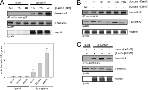
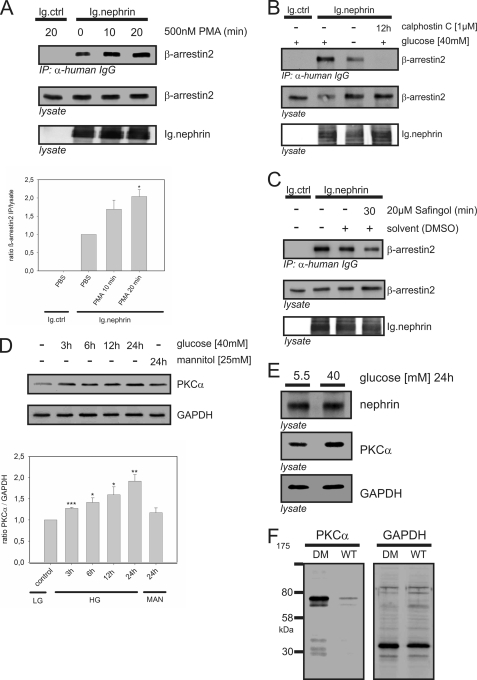
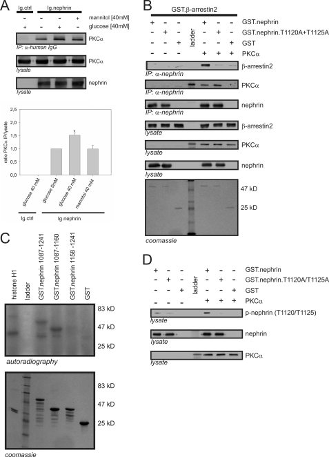
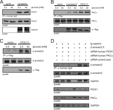
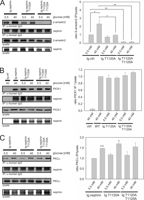
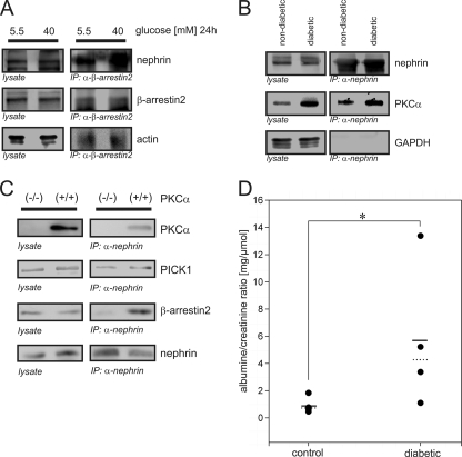
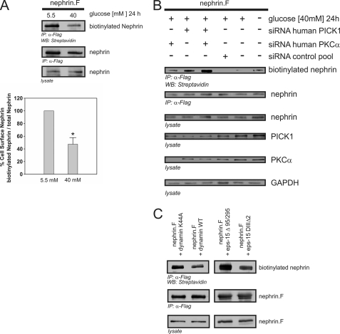
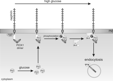
Nephrin, the key molecule of the glomerular slit diaphragm, is expressed on the surface of podocytes and is critical in preventing albuminuria. In diabetes, hyperglycemia leads to the loss of surface expression of nephrin and causes albuminuria. Here, we report a mechanism that can explain this phenomenon: hyperglycemia directly enhances the rate of nephrin endocytosis via regulation of the β-arrestin2-nephrin interaction by PKCα. We identified PKCα and protein interacting with c kinase-1 (PICK1) as nephrin-binding proteins. Hyperglycemia induced up-regulation of PKCα and led to the formation of a complex of nephrin, PKCα, PICK1, and β-arrestin2 in vitro and in vivo. Binding of β-arrestin2 to the nephrin intracellular domain depended on phosphorylation of nephrin threonine residues 1120 and 1125 by PKCα. Further, cellular knockdown of PKCα and/or PICK1 attenuated the nephrin-β-arrestin2 interaction and abrogated the amplifying effect of high blood glucose on nephrin endocytosis. In C57BL/6 mice, hyperglycemia over 24 h caused a significant increase in urinary albumin excretion, supporting the concept of the rapid impact of hyperglycemia on glomerular permselectivity. In summary, we have provided a molecular model of hyperglycemia-induced nephrin endocytosis and subsequent proteinuria and highlighted PKCα and PICK1 as promising therapeutic targets for diabetic nephropathy.


Figures

FIGURE 1.
FIGURE 1.
High glucose concentrations increase the binding of β-arrestin2 to nephrin. Coimmunoprecipitation experiments were conducted in HEK293T cells and analyzed by Western blotting. FLAG-tagged β-arrestin2 (β-arrestin2), Ig-tagged nephrin C terminus (Ig.nephrin), and Ig tag as the negative control (Ig.ctrl) were overexpressed as indicated. Staining of β-arrestin2 and nephrin in the lysate served as the internal loading control. A, impact of rising glucose concentration. HEK293T cells were incubated in the medium with increasing concentrations of glucose (5.5, 25, or 40 mm) for 24 h. After the indicated time points, coimmunoprecipitation with Ig.nephrin or Ig.ctrl was performed. Interaction was determined by staining of β-arrestin2. The results of three independent experiments were quantified by densitometry and graphed as the ratio of the β-arrestin2 immunoprecipitation (IP) signal intensity to the lysate signal intensity (ratio β-arrestin2 IP/lysate). The data are the means ± S.E. *, p < 0.05; **, p < 0.01 (Student's t test). B, time course. The cells were incubated with 5.5 mm glucose for 24 h or 40 mm glucose for 1–24 h. Then coimmunoprecipitation with Ig.nephrin or Ig.ctrl was performed. The degree of interaction was determined by staining of β-arrestin2. C, osmotic control. The cells were incubated with 5.5 or 40 mm glucose or 40 mm mannitol for 24 h. Then coimmunoprecipitation with Ig.nephrin or Ig.ctrl was performed. The degree of interaction was determined by staining of β-arrestin2.
FIGURE 2.
FIGURE 2.
PKCα mediates the high glucose effect on the nephrin-β-arrestin2 interaction. A–C, Western blots showing coimmunoprecipitation in HEK293T cells. FLAG-tagged β-arrestin2 (β-arrestin2), Ig-tagged nephrin C terminus (Ig.nephrin), and Ig tag as the negative control (Ig.ctrl) were transiently overexpressed as indicated. Staining of β-arrestin2 and nephrin in the lysate served as the internal loading control. A, impact of PKC activation on the β-arrestin2-nephrin interaction. HEK293T cells were pretreated with solvent or the general PKC activator phorbol 12-myristate 13-acetate (500 nm) for 10 or 20 min. Then coimmunoprecipitation with Ig.nephrin or Ig.ctrl was performed. The strength of interaction was determined by staining of β-arrestin2. The results of three independent experiments were quantified by densitometry and graphed as the ratio of the β-arrestin2 immunoprecipitation (IP) signal intensity to the lysate signal intensity (ratio β-arrestin2 immunoprecipitation/lysate). The data are the means ± S.E. *, p < 0.05 (Student's t test). B, impact of PKC inhibition on the β-arrestin2-nephrin interaction. HEK293T cells were maintained in 5 or 40 mm glucose and then treated with solvent or the general PKC inhibitor calphostin (1 μm) for 12 h. Coimmunoprecipitation with Ig.nephrin or Ig.ctrl was performed. The degree of interaction was measured by staining of β-arrestin2. C, isolation of the critical PKC isoform for the β-arrestin2-nephrin interaction. HEK293T cells were pretreated with solvent or the PKCα inhibitor safingol (20 μm) for 30 min. Then coimmunoprecipitation with Ig.nephrin or Ig.ctrl was performed. The degree of interaction was determined by staining of β-arrestin2. D, level of PKCα expression in immortalized podocytes under high glucose conditions. Murine podocytes were incubated in 40 mm glucose, and the PKCα protein levels were measured at indicated time points. GAPDH levels served as the internal loading control, and 40 mm mannitol (24 h) was used as the isosmotic control. The results of three independent experiments were quantified by densitometry and graphed as the ratio of the PKCα signal intensity to the GAPDH immunostaining signal intensity (ratio PKCα/GAPDH). The data are the means ± S.E. *, p < 0.05; **, p < 0.01; ***, p < 0.001 (Student's t test). E, level of PKCα expression in murine glomeruli under high glucose conditions. Isolated murine glomeruli (C57BL/6 mice) were incubated in 5.5 or 40 mm glucose for 24 h. The level of PKCα expression was determined by staining with a specific antibody. Staining of nephrin and GAPDH served as the internal loading controls. F, level of PKCα expression in the kidneys of diabetic mice. Diabetes was induced in C57BL/6 mice by injection of streptozotocin. After 24 h of glucose levels >33 mmol/liter, the kidneys were harvested and lysed, and the PKCα levels were measured. Staining of GAPDH served as the internal loading control. DM, diabetic mice.
FIGURE 3.
FIGURE 3.
Interaction of β-arrestin2 with nephrin in vitro depends on PKCα phosphorylation of nephrin threonine residues 1120 and 1125. A, Western blots showing coimmunoprecipitation in HEK293T cells overexpressing untagged PKCα (PKCα), Ig-tagged nephrin C terminus (Ig.nephrin), or Ig tag as the negative control (Ig.ctrl). The cells were maintained in 5.5 or 40 mm glucose or 40 mm mannitol (osmotic control) for 24 h. Then coimmunoprecipitation with Ig.ctrl or Ig.nephrin was performed. The level of interaction was determined by staining of PKCα. The results of three independent experiments were quantified by densitometry and graphed as the ratio of the PKCα (immunoprecipitation, IP) signal intensity to the lysate signal intensity (ratio PKCα immunoprecipitation/lysate). The data are the means ± S.E. *, p < 0.05 (Student's t test. B, in vitro pull-down assay. Aliquots of recombinant nonmutated nephrin cytoplasmic domain (GST.nephrin), double-mutant cytoplasmic domain (GST.nephrin.T1120A+T1125A), and control protein (GST) expressed in E. coli were immobilized by anti-nephrin antibody in the presence or absence of PKCα. In the Western blots, the interaction of nephrin with β-arrestin2 and PKCα was determined by staining with specific antibodies. Lysate controls show protein input of β-arrestin2, PKCα, and nephrin. Coomassie staining of SDS-PAGE gel showed equal protein input of Ig.nephrin (51 kDa) and GST (25 kDa). The molecular mass markers are indicated in kDa. C, in vitro phosphorylation assay. Aliquots of recombinant wild-type nephrin cytoplasmic domain (amino acids 1087–1241) and truncated nephrin cytoplasmic domain with (amino acids 1087–1169) and without the predicted β-arrestin2 interaction site (amino acids 1158–1241) were expressed in E. coli. Then recombinant PKCα and γ-32P were added. Histone H1 was used as the positive control. Phosphorylation was visualized by autoradiography. Coomassie staining of an SDS-PAGE gel showed equal protein input. Molecular mass markers are indicated in kDa. D, in vitro phosphorylation assay with phospho-nephrin antibody. Aliquots of recombinant nonmutated nephrin cytoplasmic domain (GST.nephrin), double-mutant cytoplasmic domain (GST.neprin.T1120A+T1125A), and control protein (GST) expressed in E. coli were immobilized by anti-nephrin antibody in the presence or absence of PKCα. In the Western blots, phosphorylation of nephrin, the mutant, and the control was visualized by staining with phospho-nephrin antibody to recognize phosphorylated nephrin threonine residues 1120 or 1125. Staining of total nephrin and PKCα served as the internal loading control.
FIGURE 4.
FIGURE 4.
Nephrin, PICK1, PKCα, and β-arrestin2 form a protein complex. A–D, Western blots showing coimmunoprecipitation (IP) in HEK293T cells overexpressing FLAG-tagged PICK1 (PICK1.F), untagged PKCα (PKCα), FLAG-tagged β-arrestin2 (β-arrestin2.F), Ig-tagged nephrin C terminus (Ig.nephrin), or Ig tag as the negative control (Ig.ctrl). A, interaction of nephrin and PICK1. HEK293T cells were incubated with 5.5 or 40 mm glucose for 24 h. Then coimmunoprecipitation with Ig.ctrl or Ig.nephrin was performed, and the interaction was determined by staining of PICK1. Staining of nephrin and PICK1 in the lysates served as the internal loading control. B, interactions of β-arrestin2 with PKCα and PICK with PKCα. HEK293T cells were incubated with 5.5 or 40 mm glucose for 24 h. Then coimmunoprecipitation with vector control, β-arrestin2, or PICK1 was performed, and the interaction was determined by staining of PKCα. Staining of PKCα and nephrin in the lysates served as the internal loading control. C, interaction of PICK1 and β-arrestin2. HEK293T cells were incubated with 5.5 or 40 mm glucose for 24 h. Then coimmunoprecipitation with vector control or PICK1 was performed, and the interaction was determined by staining of β-arrestin2. Staining of β-arrestin2 and nephrin in the lysates served as the internal loading control. D, impact of cellular knockdown of PICK1 and PKCα on β-arrestin2. HEK293T cells were transfected with Ig.nephrin and β-arrestin2. Specific siRNA was used to deplete HEK293T cells of endogenous PKCα and/or PICK1. Nephrin and β-arrestin2 were overexpressed, and nephrin was immunoprecipitated under high glucose conditions (40 mm). The β-arrestin2-nephrin interaction was determined by staining of β-arrestin2 (first row). Expression levels of β-arrestin2 (second row), nephrin (third row), PICK1 (fourth row), PKCα (fifth row), and GAPDH (sixth row) were determined by staining with specific antibodies. siRNA was transfected as follows: first column, PKCα siRNA; second column, PICK1 siRNA; third column, PKCα and PICK1 siRNA. For the controls: fourth column, endogenous protein levels, no nephrin transfected; fifth column, no siRNA, transfection lipid only; sixth column, scrambled siRNA. GAPDH was used as the internal loading control.
FIGURE 5.
FIGURE 5.
The role of nephrin threonine residues 1120 and 1125 in the protein complex. A–C, Western blots showing coimmunoprecipitation in HEK293T cells transfected with nephrin C terminus (Ig.nephrin) or Ig tag as the negative control (Ig.ctrl); single-mutant nephrin T1120A (Ig.nephrinT1120A) or double-mutant nephrin T1120A+T1125A (Ig.nephrin T1120A T1125A); and FLAG-tagged β-arrestin2 (β-arrestin2), PICK1 (PICK), or PKCα in the indicated combinations. A, β-arrestin2 binding to mutated nephrin. HEK293T cells were incubated with 5.5 or 40 mm glucose for 24 h. Then coimmunoprecipitation with Ig.nephrin, Ig.nephrinT1120A, or Ig.nephrinT1120A+T1125A was performed, and the interaction was determined by staining of β-arrestin2. Staining of β-arrestin2 and nephrin served as the internal loading control. The results of three independent experiments were quantified by densitometry and graphed as the ratio of the β-arrestin2 immunoprecipitation (IP) signal intensity to the lysate signal intensity (ratio β-arrestin2 immunoprecipitation/lysate). The data are the means ± S.E. *, p < 0.05; **, p < 0.01; ***, p < 0.001 (Student's t test). B, PICK1 binding to mutated nephrin. HEK293T cells were incubated with 5.5 or 40 mm glucose for 24 h. Then coimmunoprecipitation with Ig.nephrin, Ig.nephrinT1120A, or Ig.nephrinT1120A+T1125A was performed, and the interaction was determined by staining of PICK1. Staining of PICK1 and nephrin served as the internal loading control. The results of three independent experiments were quantified by densitometry and graphed as the ratio of the PICK1 IP signal intensity to the lysate signal intensity (ratio PICK1 IP/lysate). The data are the means ± S.E. (no significance was reached: p > 0.05). C, PKCα binding to mutated nephrin. HEK293T cells were incubated with 5.5 or 40 mm glucose for 24 h. Then coimmunoprecipitation with Ig.nephrin, Ig.nephrinT1120A, or Ig.nephrinT1120A+T1125A was performed, and the interaction was determined by staining of PKCα. Staining of PKCα and nephrin served as the internal loading control. The results of three independent experiments were quantified by densitometry and graphed as the ratio of the PKCα IP signal intensity to the lysate signal intensity (ratio PKCα IP/lysate). The data are the means ± S.E. *, p < 0.05; ***, p < 0.001 (Student's t test).
FIGURE 6.
FIGURE 6.
High glucose concentrations increase the β-arrestin2-nephrin interaction in vivo and cause albuminuria in mice. A–C, Western blots showing endogenous coimmunoprecipitations. A, endogenous interaction of β-arrestin2 and nephrin. Murine glomeruli (C57BL/6 mice) were incubated for 24 h after isolation in 5.5 or 40 mm glucose. β-Arrestin2 was immunoprecipitated, and the interaction was determined by staining of nephrin. Staining of actin served as the internal control. B, endogenous interaction of nephrin and PKCα. Diabetes was induced in C57BL/6 mice by injection of streptozotocin. After 24 h of glucose levels > 33 mmol/liter, glomeruli were isolated and lysed. Nephrin was immunoprecipitated, and the interaction was determined by staining of PKCα. Staining of immunoprecipitation and lysates of GAPDH served as the internal control. C, endogenous immunoprecipitation in murine PKCα-deficient podocytes (PKCα−/−) and wild-type control cells (PKCα+/+). Endogenous nephrin was immunoprecipitated, and the interaction of PKCα, PICK1, and β-arrestin2 was determined by staining with specific antibodies. Staining of lysates served as the internal control. D, albuminuria in diabetic versus nondiabetic C57BL/6 mice. Diabetes was induced in C57BL/6 mice by injection of streptozotocin. After 24 h of glucose levels of >33 mmol/liter, albuminuria was quantified as the albumin/creatinine ratio. Left column, nondiabetic control animals (n = 5); right column, diabetic animals (n = 4). The solid line represents the mean albumin/creatinine ratio, and the dashed line represents the median albumin/creatinine ratio values. *, p < 0.05 (Wilcoxon signed-rank test).
FIGURE 7.
FIGURE 7.
High glucose-induced nephrin endocytosis depends on PKCα and PICK1. A–C, surface abundance of nephrin measured in biotinylation assays. FLAG-tagged full-length nephrin (nephrin.F), functional dynamin (dynamin WT), and epsin (eps-15Δ95/295), and the according dominant negative mutants (dynamin K44A and eps-15 DIIIΔ2) were transiently overexpressed in HEK293T cells. A, impact of high glucose concentrations on nephrin surface abundance. After immunoprecipitation, surface nephrin was determined by detecting the biotinylated fraction. Staining of nephrin in the lysate and immunoprecipitation served as the internal control. The results were quantified by densitometry and graphed as the ratio of the PKCα immunoprecipitation (IP) signal intensity to the lysate signal intensity (ratio PKCα IP/lysate). The data are the means ± S.E. (n = 4). *, p < 0.001 (Student's t test). B, impact of cellular knockdown of PICK1 and PKCα on nephrin surface abundance. Specific siRNA was used to deplete HEK293T cells of endogenous PKCα and/or PICK1. The cells were maintained in 5.5 or 40 mm glucose medium for 24 h. GAPDH was used as the internal loading control. First column, PKCα siRNA; second column, PICK1 siRNA; third column, PKCα and PICK1 siRNA. For the controls: fourth column, scrambled control siRNA; fifth column, no siRNA (40 mm glucose); sixth column, no siRNA, transfection lipid only (5.5 mm glucose). Biotinylated surface nephrin was stained with streptavidin (first row). Total expression levels of nephrin are shown in the second row (immunoprecipitation, IP) and the third row (lysate). The expression levels of PICK1, PKCα, and GAPDH are shown in the fourth, fifth, and sixth rows, respectively. C, nephrin endocytosis depends on dynamin and clathrin. FLAG-tagged full-length nephrin (nephrin.F), dynamin (wild-type and dominant negative mutant K44A), and epsin (eps-15Δ95/295 and dominant negative mutant eps-15 DIIIΔ2) were transiently overexpressed in HEK293T cells. After immunoprecipitation, surface nephrin was determined by detecting the biotinylated fraction. Staining of nephrin in the immunoprecipitation and lysate served as the internal loading control.
FIGURE 8.
FIGURE 8.
Molecular mechanism of high glucose-induced nephrin endocytosis: hyperglycemia induces up-regulation of PKCα expression. PKCα and PICK1 are recruited to the nephrin C terminus. PKCα-mediated phosphorylation facilitates the interaction of β-arrestin2 with nephrin. β-Arrestin2 then connects nephrin to the endocytotic machinery and triggers internalization of the protein complex. Increased endocytosis leads to less surface abundance of nephrin.

References

    1. Rossing P. (2006) Curr. Diab. Rep. 6, 479–483 - PubMed
    1. Ziyadeh F. N., Wolf G. (2008) Curr. Diabetes Rev. 4, 39–45 - PubMed
    1. Kalaitzidis R., Bakris G. (2009) J. Clin. Hypertens. 11, 636–643 - PMC - PubMed
    1. Huber T. B., Benzing T. (2005) Curr. Opin. Nephrol. Hypertens. 14, 211–216 - PubMed
    1. Tryggvason K., Patrakka J., Wartiovaara J. (2006) N. Engl. J. Med. 354, 1387–1401 - PubMed

Publication types

MeSH terms