. 2011 Mar 8;108(10):4099-104.
doi: 10.1073/pnas.1014489108. Epub 2011 Feb 14.

Wounding enhances epidermal tumorigenesis by recruiting hair follicle keratinocytes

Affiliations

Wounding enhances epidermal tumorigenesis by recruiting hair follicle keratinocytes

Maria Kasper et al. Proc Natl Acad Sci U S A. .

Erratum in

  • Proc Natl Acad Sci U S A. 2012 Apr 3;109(14):5548. Svärd, Jessica [added]; Teglund, Stephan [added]

Abstract

Chronic wounds and acute trauma constitute well-established risk factors for development of epithelial-derived skin tumors, although the underlying mechanisms are largely unknown. Basal cell carcinomas (BCCs) are the most common skin cancers displaying a number of features reminiscent of hair follicle (HF)-derived cells and are dependent on deregulated Hedgehog (Hh)/GLI signaling. Here we show, in a mouse model conditionally expressing GLI1 and in a model with homozygous inactivation of Ptch1, mimicking the situation in human BCCs, that the wound environment accelerates the initiation frequency and growth of BCC-like lesions. Lineage tracing reveals that both oncogene activation and wounding induce emigration of keratinocytes residing in the lower bulge and the nonpermanent part of the HFs toward the interfollicular epidermis (IFE). However, only oncogene activation in combination with a wound environment enables the participation of such cells in the initiation of BCC-like lesions at the HF openings and in the IFE. We conclude that, in addition to the direct enhancement of BCC growth, the tumor-promoting effect of the wound environment is due to recruitment of tumor-initiating cells originating from the neighboring HFs, establishing a link between epidermal wounds and skin cancer risk.


Conflict of interest statement

The authors declare no conflict of interest.

Figures
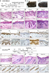
Fig. 1.
Fig. 1.
Postnatal activation of Hh signaling induces formation of BCC-like lesions in mice. (A) Experimental timeline for GLI1 overexpression experiments. (B) Standing hair and hyperkeratosis in the dorsal skin of a K5tTA/TREGLI1 (K5/GLI1) mouse. (C–F) H&E stained dorsal skin sections from K5tTA/TREGLI1 mice reflecting the stages of tumor progression from small IFE-associated proliferations to more advanced BCC-like lesions originating from both IFE and HFs. (G and H) Immunostaining for marker gene expression in stage 2 (G) and stage 3 (H) lesions illustrates expression of K5, Sox9, and P-cadherin characteristic for basaloid proliferations. Note that the lesions are K6-negative at the protruding front. (I–L) Stages of tumor progression in K5Cre*PR1/Ptch1fl/fl mice after inactivation of both Ptch1 alleles. (I) Experimental timeline for Ptch1 deletion. (J) Stage 1 lesions arising from HFs in dorsal skin. (K) Proliferations in IFE and HF in stage 2 dorsal skin. (L) Stage 3 lesions in ear skin from the mice shown in Fig. 1K. (M) Immunostaining for marker gene expression in stage 1 lesions. (C–F and J–L) H&E staining. (G, H, and M) Hematoxylin counterstain. (Scale bars: C–E Upper, F, and J–M, 100 μm; C–E Lower, G, and H, 50 μm.)
Fig. 2.
Fig. 2.
Wounding enhances the formation of BCC-like lesions. (A) IFE-associated lesions in unwounded skin of K5tTA/TREGLI1 (K5/GLI1) mice: small, stage 2 buds are indicated by arrowheads. (B) Lesions developing in the wound epidermis of the same mouse were more advanced, classified as stage 3 lesions, and were also found at HF openings. (C) No lesions developed in the wound epidermis of TREGLI1 mice. (D) Quantification of GLI1-induced lesions in unwounded and wounded skin showed that the number of lesions remained constant, whereas the size of the lesions was significantly increased in the wound area (**P = 0.02; n = 3; error bars indicate SD). (E) Immunostaining for marker gene expression of lesions in the wound epidermis shows the typical staining pattern for basaloid lesions. (F) Small, stage 2 lesions in unwounded skin of K5Cre*PR1/Ptch1fl/fl mice associated with HF or IFE (arrowheads). (G) Enhanced formation of lesions at the site of wounding in K5Cre*PR1/Ptch1fl/fl epidermis (arrowheads). (H) In the wound epidermis of K5Cre*PR1/Ptch1+/fl, no lesions formed. (I) Quantification of the lesions revealed a significant increase in the overall number and the average size of the proliferations in the wounded skin compared with unwounded skin of K5Cre*PR1/Ptch1fl/fl mice (**P = 0.01; ***P = 0.001; n = 3; error bars indicate SD). (J) Characteristic staining of lesions in the wound epidermis for basaloid proliferation markers confirms their identity as evolving BCC-like lesions. (A–C and F–H) H&E staining. (E and J) Hematoxylin counterstain. (Scale bars: A–C and F–H, 100 μm; E and J, 50 μm.)
Fig. 3.
Fig. 3.
Keratinocytes from the bulge and the nonpermanent part of the HF contribute to tumor formation in the IFE only in the wound area. (A) Experimental timeline of Lgr5+ lineage tracing in Lgr5-EGFP-IRES-creERT2/R26R mice. Tamoxifen (TM) was administered at P14, wounds were created at 5 wk of age, and samples were taken at 7 wk of age. (B) LacZ staining of Lgr5-EGFP-IRES-creERT2/R26R normal skin at 7 wk of age. Only the part of the HF extending up to the level of the sebaceous gland (SG) opening (dashed line) was repopulated by Lgr5+ progeny. (C and D) 2 wk after wounding, Lgr5+ progeny had advanced to the permanent and infundibular parts of the HF located close to the wound. Dashed line indicates level of SG openings (C). In addition, Lgr5+ progeny were integrated into the newly formed wound epidermis (D). (E) Experimental timeline of Lgr5+ tracing with subsequent tumor induction in K5tTA/TREGLI1/Lgr5-EGFP-IRES-creERT2/R26R mice. (F) At 5 wk of age, when stage 1 proliferations appear in the IFE (Inset, arrowhead), the progeny of Lgr5+ cells is located below the SG openings (dashed line). (G) As the tumors in the IFE progress to stage 2 (arrowhead), the labeled Lgr5+ progeny advances to the HF infundibulum. (H) Concomitantly with the appearance of the stage 3 lesions (arrowhead), the Lgr5+ progeny move further away from the HF and differentiate but do not integrate into the basaloid lesions in the IFE. Arrow, HF opening. (I) Experimental timeline of Lgr5+ tracing in combined tumor induction and wounding experiments. (J) Lgr5+ progeny can integrate in the wound epidermis in the presence of activated GLI1 expression. (K) The Lgr5+ progeny participate in the formation of K5-positive buds (L), which can be identified as early basaloid lesions based on their positive immunostaining for Sox9. (B–D and F Inset) LacZ and H&E staining. (F–H and J) LacZ and eosin staining. (K and L) Hematoxylin counterstain. SHG, secondary hair germ. (Scale bars: B, C, F–H, and J, 100 μm; D, K, and L, 50 μm.)
Fig. 4.
Fig. 4.
Lgr5+ keratinocytes represent tumor-initiating cells for HF-associated lesions and for IFE-associated lesions after wounding. (A) Tamoxifen-injected Lgr5-EGFP-IRES-creERT2/Ptch1+/fl control mice show no phenotype in unwounded skin. (B) Basaloid proliferations in unwounded dorsal skin of 2-mo-old Lgr5-EGFP-IRES-creERT2/Ptch1fl/fl mice. (C) HF-associated BCC-like lesions in the unwounded dorsal skin of a 4-mo-old Lgr5-EGFP-IRES-creERT2/Ptch1fl/fl mouse. (Left) H&E staining. (Right) K6 immunostaining. (D) Dorsal wound areas of tamoxifen-injected Lgr5-EGFP-IRES-creERT2/Ptch1+/fl control mice are devoid of basaloid lesions. (E and F) The wound areas of mice shown in Fig. 4 B and C contain lesions associated with the infundibulum of the HFs as well as IFE-associated lesions. (G) Immunostaining of marker genes identifies the infundibular- and IFE-associated lesions as basaloid lesions. Arrowheads show early infundibular and IFE-associated BCC-like lesions. (A–F) H&E staining. (C Right and G) Hematoxylin counterstain. (Scale bars: 100 μm.)
Fig. 5.
Fig. 5.
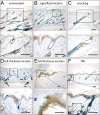
Full-thickness cutaneous wounding is required to induce HF keratinocyte migration to the IFE. (A) Lgr5+ lineage tracing in untreated Lgr5-EGFP-IRES-creERT2/R26R mice. (B–F) Tamoxifen was administered at P14; trauma was introduced on dorsal telogen skin. The samples were collected 7 d after trauma (B–E) and 14 d after repeated TPA treatment (F). (B) Superficial incisions induced K6 expression in the regenerating IFE but did not induce keratinocyte migration from the HF. (C) Hair plucking induced anagen in the affected HFs without inducing keratinocyte migration into the IFE. (D and E) Full-thickness incisions and excisional wounds induced integration of cells originating from the bulge and the nonpermanent part of the HF into the IFE. The incisional wound areas were identified by K6 staining in the IFE and the damaged muscle layer (arrows). (F) Repeated TPA treatment twice a week for 2 wk induced a hyperproliferative state in the IFE and emigration of Lgr5+ progeny into the IFE. (Inset) Non–TPA-treated skin of the same animal. (A–F) LacZ staining, K6 immunostaining, hematoxylin counterstain. (Scale bars: 100 μm.)

Comment in

  • Tumorigenesis: Wound-up tumours.
    Villanueva T. Villanueva T. Nat Rev Cancer. 2011 Apr;11(4):235. doi: 10.1038/nrc3043. Nat Rev Cancer. 2011. PMID: 21548398 No abstract available.

References

    1. Jaks V, Kasper M, Toftgård R. The hair follicle—a stem cell zoo. Exp Cell Res. 2010;316:1422–1428. - PubMed
    1. Morris RJ, et al. Capturing and profiling adult hair follicle stem cells. Nat Biotechnol. 2004;22:411–417. - PubMed
    1. Snippert HJ, et al. Lgr6 marks stem cells in the hair follicle that generate all cell lineages of the skin. Science. 2010;327:1385–1389. - PubMed
    1. Jensen KB, et al. Lrig1 expression defines a distinct multipotent stem cell population in mammalian epidermis. Cell Stem Cell. 2009;4:427–439. - PMC - PubMed
    1. Jaks V, et al. Lgr5 marks cycling, yet long-lived, hair follicle stem cells. Nat Genet. 2008;40:1291–1299. - PubMed

Publication types