. 2011 Mar 1;108(9):3671-6.
doi: 10.1073/pnas.1019423108. Epub 2011 Feb 14.

Identification of the meiotic life cycle stage of Trypanosoma brucei in the tsetse fly

Affiliations

Identification of the meiotic life cycle stage of Trypanosoma brucei in the tsetse fly

Lori Peacock et al. Proc Natl Acad Sci U S A. .

Abstract

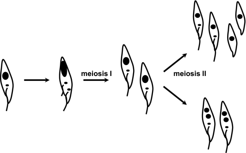
Elucidating the mechanism of genetic exchange is fundamental for understanding how genes for such traits as virulence, disease phenotype, and drug resistance are transferred between pathogen strains. Genetic exchange occurs in the parasitic protists Trypanosoma brucei, T. cruzi, and Leishmania major, but the precise cellular mechanisms are unknown, because the process has not been observed directly. Here we exploit the identification of homologs of meiotic genes in the T. brucei genome and demonstrate that three functionally distinct, meiosis-specific proteins are expressed in the nucleus of a single specific cell type, defining a previously undescribed developmental stage occurring within the tsetse fly salivary gland. Expression occurs in clonal and mixed infections, indicating that the meiotic program is an intrinsic but hitherto cryptic part of the developmental cycle of trypanosomes. In experimental crosses, expression of meiosis-specific proteins usually occurred before cell fusion. This is evidence of conventional meiotic division in an excavate protist, and the functional conservation of the meiotic machinery in these divergent organisms underlines the ubiquity and basal evolution of meiosis in eukaryotes.


Conflict of interest statement

The authors declare no conflict of interest.

Figures

Fig. 1.
Fig. 1.
Expression of meiosis-specific YFP fusion proteins in trypanosomes from SGs of tsetse flies. (A) (Top) J10 YFP::MND1. (Middle) J10 YFP::DMC1. (Bottom) J10 YFP::HOP1. The first column shows phase contrast images of fixed trypanosomes in salivary exudate; the other columns show epifluorescence microscopy images of YFP fusion protein expression or DAPI-stained nucleus and kinetoplast, along with merged images. In all cases, YFP fluorescence colocalizes with DAPI-stained nucleus toward the posterior end of the trypanosome, and there are two kinetoplasts, (center and anterior small blue dots). (B) Live phase contrast and epifluorescence images of trypanosomes of clone J10 YFP::HOP1 inside a tsetse SG. Trypanosomes expressing the fluorescent fusion protein have a blunt (asterisk) or pointed (arrow) posterior, with the nucleus very near the posterior end. The RH nonfluorescent trypanosome is a typical epimastigote with an elongated tube-like posterior. Cell movement has compromised merge of phase and fluorescence images. (Scale bar: 5 μm.)
Fig. 2.
Fig. 2.
Morphology of the meiotic cell. (A) (Top) J10 YFP::MND1, YFP::PFR1. (Middle) J10 YFP::DMC1, YFP::PFR1. (Bottom) J10 YFP::HOP1, YFP::PFR1. The first column shows phase contrast images of fixed trypanosomes in salivary exudate; the other columns show epifluorescence microscopy images of YFP fusion protein expression and merged images. In trypanosomes expressing YFP::PFR1, the PFR incorporates the fusion protein and is fluorescent. The brightly fluorescent anterior PFR is old, whereas the less-bright PFR (arrow) is that of the daughter cell. (Scale bar 5 μm.) (B) Identification of the daughter flagellum in dividing procyclic cells. Previous studies of the process of cell division and timing of construction of new organelles have shown that the daughter flagellum emerges posterior to the parental flagellum (12). In the examples shown, the new PFR (arrow) is seen to fluoresce less brightly than the old PFR in the phase contrast and epifluorescence images. (Scale bar: 5 μm.)
Fig. 3.
Fig. 3.
Comparison of morphological parameters in HMG expressers. (A) Measurements: (a) distance between the start of the PFR of each flagellum; (b) total cell length; (c) nucleus length; (d) posterior of th cell to posterior of the nucleus; (e) distance between kinetoplasts 1 and 2 (kin1–kin2); (f) distance between the nucleus and kinetoplast 1 (kin1); (g) nucleus width; (h), nucleus perimeter; (i) nucleus area. The open circle represents the nucleus; filled circles, kinetoplasts 1 and 2; thick lines, PFR. (B) Each parameter was measured in the three cell lines shown in Fig. 2. For PFR, n = 13 for MND1 and DMC1 and n = 18 for HOP1; for other parameters, n = 11 for MND1 and HOP1 and n = 14 for DMC1. P values were calculated by ANOVA and Tukey's post hoc test. *P < 0.05; **P < 0.01; ***P < 0.001. Error bars are SEM. (C) Comparative morphology of HMG expressers based on mean values (1-μm grid). The short inter-PFR distance places MND1 expressers at the start of the series; the cell length of HOP1 expressers places them at the end of the series. The nucleus progressively elongates and moves posteriorly. The relative position of the kinetoplasts remains constant.
Fig. 4.
Fig. 4.
Meiosis occurs before fusion. (A) Epifluorescence image of a section of an SG infected with J10 YFP::DMC1 and 1738 mRFP. The trypanosomes expressing YFP in the nucleus (J10 YFP::DMC1) are separate from those expressing mRFP in the cytoplasm (1738 mRFP). (B) Similar image of an SG infected with J10 YFP::HOP1 and 1738 mRFP. This rare example of a trypanosome expressing both mRFP in the cytoplasm and YFP in the nucleus suggests that in this case, fusion occurred before completion of meiosis. Live images. (Scale bar: 10 μm.)
Fig. 5.
Fig. 5.
Model of meiosis in trypanosomes. An epimastigote (Left) enters meiosis, and the first division results in two 2N cells. Meiosis II follows, producing haploid nuclei. Two possible outcomes are shown, assuming that replication of the kinetoplast and flagellum does not occur.

References

    1. Heitman J. Sexual reproduction and the evolution of microbial pathogens. Curr Biol. 2006;16:R711–R725. - PubMed
    1. Jenni L, et al. Hybrid formation between African trypanosomes during cyclical transmission. Nature. 1986;322:173–175. - PubMed
    1. Gaunt MW, et al. Mechanism of genetic exchange in American trypanosomes. Nature. 2003;421:936–939. - PubMed
    1. Akopyants NS, et al. Demonstration of genetic exchange during cyclical development of Leishmania in the sand fly vector. Science. 2009;324:265–268. - PMC - PubMed
    1. MacLeod A, et al. Allelic segregation and independent assortment in T. brucei crosses: Proof that the genetic system is Mendelian and involves meiosis. Mol Biochem Parasitol. 2005;143:12–19. - PubMed

Publication types