. 2011 Mar 1;108(9):3767-72.
doi: 10.1073/pnas.1006741108. Epub 2011 Feb 14.

Toxoplasma gondii sequesters centromeres to a specific nuclear region throughout the cell cycle

Affiliations

Toxoplasma gondii sequesters centromeres to a specific nuclear region throughout the cell cycle

Carrie F Brooks et al. Proc Natl Acad Sci U S A. .

Abstract

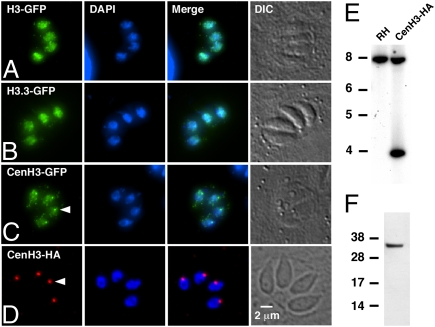
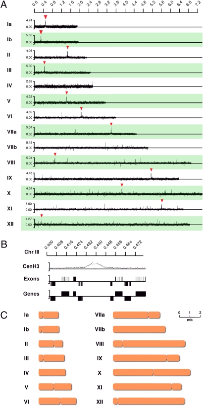
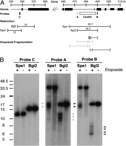
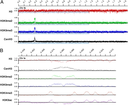
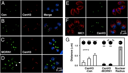
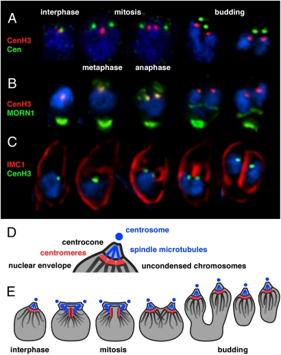
Members of the eukaryotic phylum Apicomplexa are the cause of important human diseases including malaria, toxoplasmosis, and cryptosporidiosis. These obligate intracellular parasites produce new invasive stages through a complex budding process. The budding cycle is remarkably flexible and can produce varied numbers of progeny to adapt to different host-cell niches. How this complex process is coordinated remains poorly understood. Using Toxoplasma gondii as a genetic model, we show that a key element to this coordination is the centrocone, a unique elaboration of the nuclear envelope that houses the mitotic spindle. Exploiting transgenic parasite lines expressing epitope-tagged centromeric H3 variant CenH3, we identify the centromeres of T. gondii chromosomes by hybridization of chromatin immunoprecipitations to genome-wide microarrays (ChIP-chip). We demonstrate that centromere attachment to the centrocone persists throughout the parasite cell cycle and that centromeres localize to a single apical region within the nucleus. Centromere sequestration provides a mechanism for the organization of the Toxoplasma nucleus and the maintenance of genome integrity.


Conflict of interest statement

The authors declare no conflict of interest.

Figures

Fig. 1.
Fig. 1.
T. gondii histone H3 variants. (AC) Immunofluorescence assays of tachyzoites transiently transfected with constructs expressing TGME49_061240 (H3) (A), TGME49_018260 (H3.3) (B), and TGME49_025410 (CenH3) (C) fused to the N terminus of YFP. Anti-GFP antibody is shown in green; DAPI is shown in blue. (D) To construct a strain carrying an epitope tag in the genomic locus of CenH3, a suitable cosmid was engineered by recombination in EL250 cells and transfected into T. gondii. These TgCenH3-HA lines showed a single spot of nuclear staining (red, arrowhead) in the majority of parasites in immunofluorescence. (E) Southern analysis of a stable TgCenH3-HA clone shows the presence of two bands when probed with the CenH3 coding sequence, suggesting nonhomologous insertion. (F) This clone expressed a protein of the expected size when probed in Western blot.
Fig. 2.
Fig. 2.
ChIP of CenH3-associated DNA and microarray analysis. (A) ChIP-chip analysis of DNA isolated from the transgenic CenH3-HA strain using an HA epitope-specific antibody. All 14 T. gondii chromosomes are shown. Significant hybridization peaks are indicated by red arrowheads. The x axis shows the position of probes along the chromosome; the y axis shows relative fluorescence intensity for each oligonucleotide. (B) Representative detailed view of CenH3-associated regions on chromosome III. Gene models and exon predictions for this region (version 4 annotation; www.toxodb.org) are shown below (up is sense; down is antisense). Note that CenH3-associated DNA is devoid of protein-coding genes. (C) Schematic representation of the positions of the centromeres of 12 of the 14 T. gondii chromosomes.
Fig. 3.
Fig. 3.
Etoposide mapping of the centromere of chromosome Ia. (A) Schematic map of chromosome Ia showing the CenH3-associated region as well as a control region located ∼115 kb 5′. Position of restriction fragments, probes, and mapped etoposide-induced fragments are indicated. (B) T. gondii-infected cultures were incubated for 2 h in medium containing 100 μM etoposide (+) or DMSO solvent as control (−). Parasites were purified, and genomic DNA was extracted, restricted with the indicated enzymes, and subjected to Southern analysis using the indicated probes. Note etoposide-induced fragmentation of the putative centromeric region [probes A (light gray arrowheads) and B (dark gray arrowheads)], which was not observed for the control regions (probe C).
Fig. 4.
Fig. 4.
Histone H3K9 di- and trimethylation marks the boundaries of the T. gondii centromeres. ChIP-chip analysis using the indicated antibodies was conducted as detailed in Materials and Methods. (A) Full view of chromosome Ib. Note that centromeric H3K9me2 and H3K9me3 labeling represents the most significant accumulation of these modifications across the T. gondii chromosome. (B) Detailed view of the centromeric region of chromosome Ia. Note the absence of centromeric signal for marks previously associated with promoter regions in T. gondii [H3K4me3 and histone H3 acetylated at lysine 9 (H3K9ac) (23)].
Fig. 5.
Fig. 5.
The T. gondii centromeres remain proximal to the centrocone at all times. Host-cell cultures were infected with CenH3-HA transgenic parasites and fixed and processed for immunofluorescence 24 h later. Cells were stained with an antibody to HA (CenH3) and centrin (Cen) (A and B), MORN1 (C and D), and IMC1 (E and F). Representative images are shown for nondividing (A, C, and E) and dividing parasites (B, D, and F). The vertical arrowhead in D indicates the centrocone, and the horizontal arrowhead indicates the daughter-cell basal complex. (G) The distance from the center of CenH3 labeling to the center of the centrin or MORN1 label was measured by image analysis. The mean distances from centrin (54 nuclei) and MORN1 (70 nuclei) are shown. Error bars depict SD. Values were grouped by cell-cycle phase as indicated by symbols at the top of the graph. (See Fig. 6 for further details about T. gondii mitosis.) The radii of nuclei are shown for size comparison (area of DAPI-stained nuclei was measured and radii were calculated from area, assuming a circular shape for simplicity; n = 52 nuclei). ***P < 0.001.
Fig. 6.
Fig. 6.
Toxoplasma mitosis and budding. (A, B, and C) Immunofluorescence micrographs showing details for individual nuclei at key points of the T. gondii cell-division cycle (note that each panel represents five individual cells consolidated and cropped from different micrographs). Three-color merged images are shown for the antibodies indicated and the DNA stained with DAPI (blue). Note that MORN1 staining is present in the centrocones of all budding stages but is not visible in the two panels showing budding in B because of stronger signal from the CenH3 (red channel). (D) Schematic view of the centrocone highlighting its components. (See ref. for additional details.) (E) Model of centromere sequestration throughout the parasite cell cycle.

References

    1. Sibley LD. Intracellular parasite invasion strategies. Science. 2004;304:248–253. - PubMed
    1. Striepen B, Jordan CN, Reiff S, van Dooren GG. Building the perfect parasite: Cell division in Apicomplexa. PLoS Pathog. 2007;3:e78. - PMC - PubMed
    1. Sheffield HG, Melton ML. The fine structure and reproduction of Toxoplasma gondii. J Parasitol. 1968;54:209–226. - PubMed
    1. Bannister LH, Hopkins JM, Fowler RE, Krishna S, Mitchell GH. A brief illustrated guide to the ultrastructure of Plasmodium falciparum asexual blood stages. Parasitol Today. 2000;16:427–433. - PubMed
    1. Speer CA, Dubey JP. Ultrastructure of shizonts and merozoites of Sarcocystis falcatula in the lungs of budgerigars (Melopsittacus undulatus) J Parasitol. 1999;85:630–637. - PubMed

Publication types