. 2011 May 12;117(19):5207-14.
doi: 10.1182/blood-2010-08-302018. Epub 2011 Feb 14.

An embryonic stage-specific enhancer within the murine β-globin locus mediates domain-wide histone hyperacetylation

Affiliations

An embryonic stage-specific enhancer within the murine β-globin locus mediates domain-wide histone hyperacetylation

George Fromm et al. Blood. .

Abstract

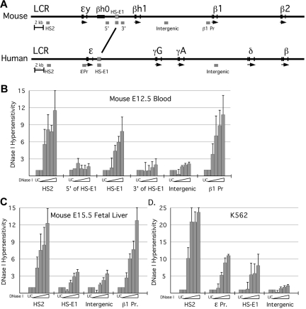
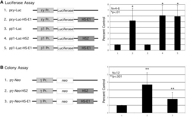
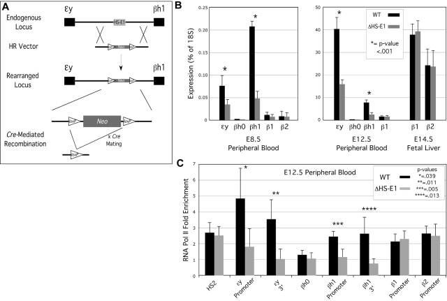
In mammalian nuclei, a select number of tissue-specific gene loci exhibit broadly distributed patterns of histone modifications, such as histone hyperacetylation, that are normally associated with active gene promoters. Previously, we characterized such hyperacetylated domains within mammalian β-globin gene loci, and determined that within the murine locus, neither the β-globin locus control region nor the gene promoters were required for domain formation. Here, we identify a developmentally specific erythroid enhancer, hypersensitive site-embryonic 1 (HS-E1), located within the embryonic β-globin domain in mouse, which is homologous to a region located downstream of the human embryonic ε-globin gene. This sequence exhibits nuclease hypersensitivity in primitive erythroid cells and acts as an enhancer in gain-of-function assays. Deletion of HS-E1 from the endogenous murine β-globin locus results in significant decrease in the expression of the embryonic β-globin genes and loss of the domain-wide pattern of histone hyperacetylation. The data suggest that HS-E1 is an enhancer that is uniquely required for β-like globin expression in primitive erythroid cells, and that it defines a novel class of enhancer that works in part by domain-wide modulation of chromatin structure.


Figures

Figure 1
Figure 1
DNase I HS analysis in erythroid cells. (A) Schematic of the mouse and human β-globin loci, to scale. Positions of PCR amplimers used in the DNaseI assay are indicated as gray bars beneath each locus. The region of homology between mouse and human to which HS-E1 maps is marked by the line between the 2 loci (see also supplemental Figure 1). HS analysis was performed for the murine β-globin locus using primary erythroid cells harvested from (B) E12.5 peripheral blood and (C) E15.5 fetal liver. HS analysis was also performed for the human β-globin locus using the K562 cell line (D). Analysis was performed using qPCR and the loss in fluorescent signal indicated a decrease in amplification product. This value was interpreted as an increase in hypersensitivity, and the inverse of this value was represented heregraphically.
Figure 2
Figure 2
Assays for enhancer function of HS-E1. (A) Transient reporter assay. Reporter constructs used in the transient assay are illustrated at the left. The bar graph shows relative levels of luciferase activity measured using the indicated constructs. (B) Colony assay. The selectable marker constructs used in the colony assay are illustrated to the left. The bar graph shows colony numbers obtained using the indicated constructs, with the average colony number for pγneo normalized to 1.0. Each bar in the graph represents the average of 12 replicates of the experiment. All error bars represent standard error of the mean.
Figure 3
Figure 3
Effect of deletion of HS-E1 from the endogenous β-globin locus on gene expression and RNA Pol II association. (A) Homologous recombination strategy for the deletion of HS-E1. (B) β-globin mRNA expression analysis for E8.5 and E12.5 peripheral blood, and for E14.5 fetal liver, derived from wild-type embryos and embryos homozygous for the deletion of HS-E1. All values are derived from averages of at least 6 individual embryos. (C) RNA Pol II ChIP analysis in E12.5 peripheral blood. Enrichment for RNA polymerase II association with the indicated sequences is calculated using inactive control loci as background controls (ie, 1.0).
Figure 4
Figure 4
Effect of deletion of HS-E1 on domain-wide histone modifications. A schematic of the murine β-globin locus is shown at the top, to scale but with the indicated discontinuities. The bracketed region between the ϵy-globin and βh1-globin genes shows the location of the HS-E1 deletion. Black bars beneath the locus correspond to the positions of amplimers used for ChIP analysis, and the corresponding ChIP data are shown directly below each amplimer in the bar graphs. Values in the bar graphs represent enrichments observed for the indicated histone modifications relative to inactive control loci, as measured in E12.5 primitive erythroid cells from wild-type mice (black) or from mice homozygous for the deletion of HS-E1 (gray). Note that one amplimer lies within the deleted region.

References

    1. Felsenfeld G, Groudine M. Controlling the double helix. Nature. 2003;421(6921):448–453. - PubMed
    1. Kurdistani SK, Grunstein M. Histone acetylation and deacetylation in yeast. Nat Rev Mol Cell Biol. 2003;4(4):276–284. - PubMed
    1. Bulger M. Hyperacetylated chromatin domains: lessons from heterochromatin. J Biol Chem. 2005;280(23):21689–21692. - PubMed
    1. Fromm G, Bulger M. A spectrum of gene regulatory phenomena at mammalian beta-globin gene loci. Biochem Cell Biol. 2009;87(5):781–790. - PubMed
    1. Bender MA, Bulger M, Close J, Groudine M. Beta-globin gene switching and DNase I sensitivity of the endogenous beta-globin locus in mice do not require the locus control region. Mol Cell. 200;5(2):387–393. - PubMed

Publication types