

Excitation-resolved fluorescence tomography with simplified spherical harmonics equations
- PMID: 21321388
- PMCID: PMC3679937
- DOI: 10.1088/0031-9155/56/5/015
Excitation-resolved fluorescence tomography with simplified spherical harmonics equations
Abstract
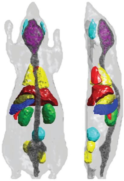
Fluorescence tomography (FT) reconstructs the three-dimensional (3D) fluorescent reporter probe distribution inside biological tissue. These probes target molecules of biological function, e.g. cell surface receptors or enzymes, and emit fluorescence light upon illumination with an external light source. The fluorescence light is detected on the tissue surface and a source reconstruction algorithm based on the simplified spherical harmonics (SP(N)) equations calculates the unknown 3D probe distribution inside tissue. While current FT approaches require multiple external sources at a defined wavelength range, the proposed FT method uses only a white light source with tunable wavelength selection for fluorescence stimulation and further exploits the spectral dependence of tissue absorption for the purpose of 3D tomographic reconstruction. We will show the feasibility of the proposed hyperspectral excitation-resolved fluorescence tomography method with experimental data. In addition, we will demonstrate the performance and limitations of such a method under ideal and controlled conditions by means of a digital mouse model and synthetic measurement data. Moreover, we will address issues regarding the required amount of wavelength intervals for fluorescent source reconstruction. We will explore the impact of assumed spatially uniform and nonuniform optical parameter maps on the accuracy of the fluorescence source reconstruction. Last, we propose a spectral re-scaling method for overcoming the observed limitations in reconstructing accurate source distributions in optically non-uniform tissue when assuming only uniform optical property maps for the source reconstruction process.
Figures


Similar articles
-
Hyperspectral excitation-resolved fluorescence tomography of quantum dots.Opt Lett. 2009 Aug 15;34(16):2477-9. doi: 10.1364/ol.34.002477. Opt Lett. 2009. PMID: 19684821 Free PMC article.
-
Excitation spectroscopy in multispectral optical fluorescence tomography: methodology, feasibility and computer simulation studies.Phys Med Biol. 2009 Aug 7;54(15):4687-704. doi: 10.1088/0031-9155/54/15/004. Epub 2009 Jul 10. Phys Med Biol. 2009. PMID: 19590118 Free PMC article.
-
Bioluminescence tomography with CT/MRI co-registration.Annu Int Conf IEEE Eng Med Biol Soc. 2009;2009:6327-30. doi: 10.1109/IEMBS.2009.5333170. Annu Int Conf IEEE Eng Med Biol Soc. 2009. PMID: 19964154
-
High-performance fluorescence molecular tomography through shape-based reconstruction using spherical harmonics parameterization.PLoS One. 2014 Apr 14;9(4):e94317. doi: 10.1371/journal.pone.0094317. eCollection 2014. PLoS One. 2014. PMID: 24732826 Free PMC article.
-
A multi-view time-domain non-contact diffuse optical tomography scanner with dual wavelength detection for intrinsic and fluorescence small animal imaging.Rev Sci Instrum. 2012 Jun;83(6):063703. doi: 10.1063/1.4726016. Rev Sci Instrum. 2012. PMID: 22755630 Review.
Cited by
-
Macroscopic-imaging technique for subsurface quantification of near-infrared markers during surgery.J Biomed Opt. 2015 Mar;20(3):036014. doi: 10.1117/1.JBO.20.3.036014. J Biomed Opt. 2015. PMID: 25793562 Free PMC article.
-
In vivo bioluminescence tomography-guided system for pancreatic cancer radiotherapy research.Biomed Opt Express. 2024 Jul 9;15(8):4525-4539. doi: 10.1364/BOE.523916. eCollection 2024 Aug 1. Biomed Opt Express. 2024. PMID: 39347008 Free PMC article.
-
Automated quantification of bioluminescence images.Nat Commun. 2018 Oct 15;9(1):4262. doi: 10.1038/s41467-018-06288-w. Nat Commun. 2018. PMID: 30323260 Free PMC article.
-
Separating structures of different fluorophore concentrations by principal component analysis on multispectral excitation-resolved fluorescence tomography images.Biomed Opt Express. 2013 Aug 29;4(10):1829-45. doi: 10.1364/BOE.4.001829. eCollection 2013. Biomed Opt Express. 2013. PMID: 24156047 Free PMC article.
-
Spectral-resolved cone-beam X-ray luminescence computed tomography with principle component analysis.Biomed Opt Express. 2018 May 30;9(6):2844-2858. doi: 10.1364/BOE.9.002844. eCollection 2018 Jun 1. Biomed Opt Express. 2018. PMID: 30258694 Free PMC article.
References
-
- Arridge SR. Optical tomography in medical imaging. Inverse Problems. 1999;15:R41–93.
-
- Aydin ED, de Oliveira CRE, Goddard AJH. A comparison between transport and diffusion calculations using a finite-element spherical harmonics radiation transport method. Med. Phys. 2002;29:2013–23. - PubMed
-
- Biju V, Itoh T, Anas A, Sujith A, Ishikawa M. Semiconductor quantum dots and metal nanoparticles: syntheses, optical properties, and biological applications. Anal. Bioanal. Chem. 2008;391:2469–95. - PubMed
Publication types
MeSH terms
Grants and funding
LinkOut - more resources
Full Text Sources
Other Literature Sources