. 2011 May;21(5):741-53.
doi: 10.1038/cr.2011.29. Epub 2011 Feb 15.

Transport of receptors, receptor signaling complexes and ion channels via neuropeptide-secretory vesicles

Affiliations

Transport of receptors, receptor signaling complexes and ion channels via neuropeptide-secretory vesicles

Bo Zhao et al. Cell Res. 2011 May.

Abstract

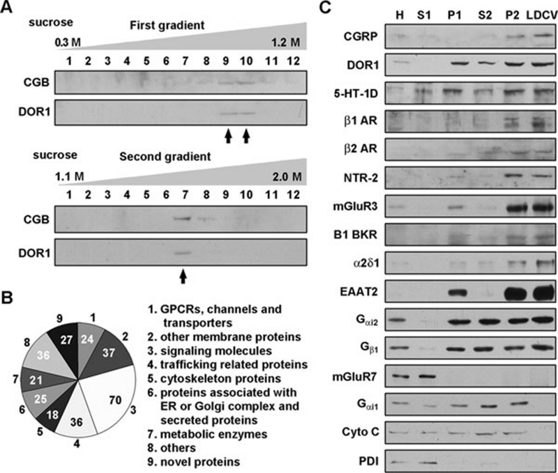
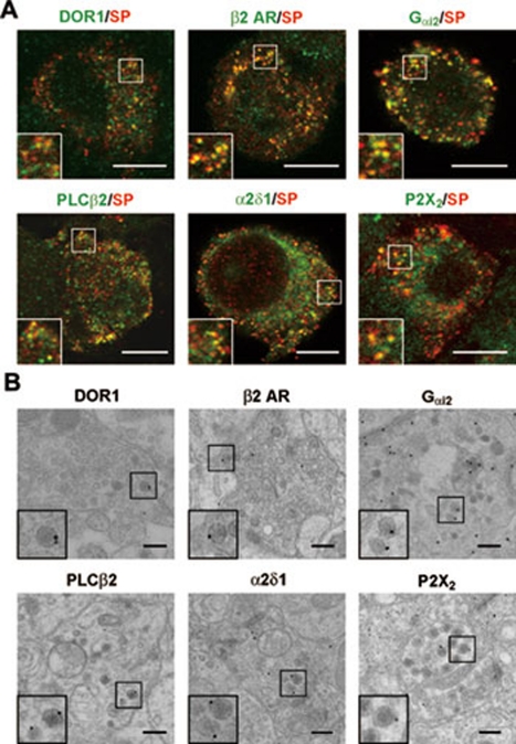
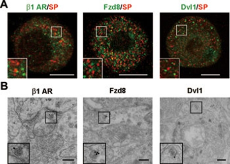
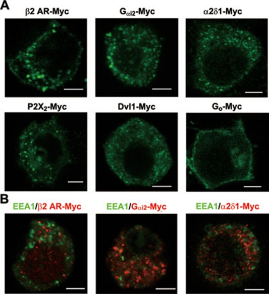
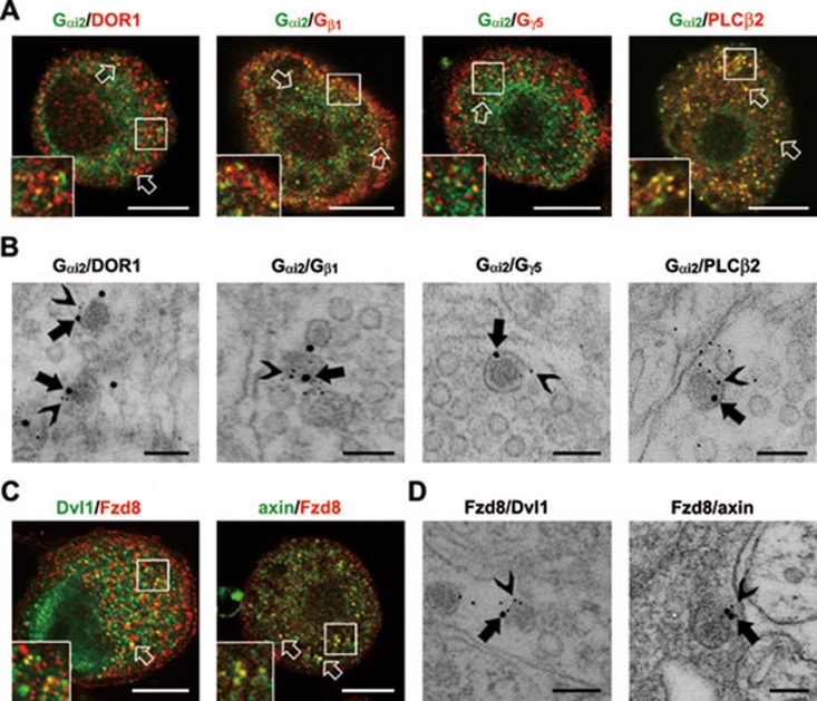
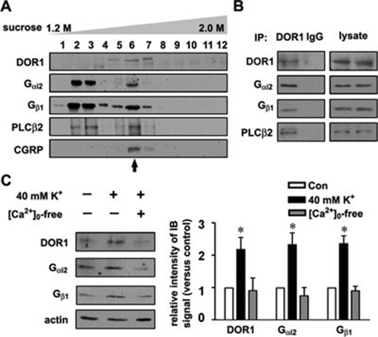
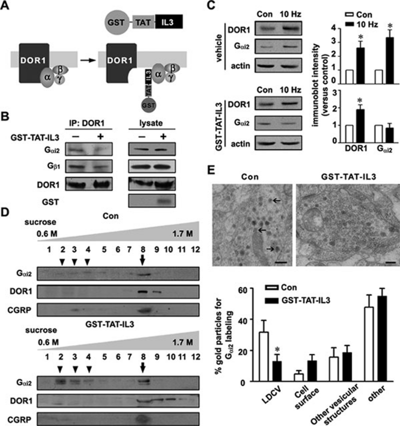
Stimulus-induced exocytosis of large dense-core vesicles (LDCVs) leads to discharge of neuropeptides and fusion of LDCV membranes with the plasma membrane. However, the contribution of LDCVs to the properties of the neuronal membrane remains largely unclear. The present study found that LDCVs were associated with multiple receptors, channels and signaling molecules, suggesting that neuronal sensitivity is modulated by an LDCV-mediated mechanism. Liquid chromatography-mass spectrometry combined with immunoblotting of subcellular fractions identified 298 proteins in LDCV membranes purified from the dorsal spinal cord, including G-protein-coupled receptors, G-proteins and other signaling molecules, ion channels and trafficking-related proteins. Morphological assays showed that δ-opioid receptor 1 (DOR1), β2 adrenergic receptor (AR), G(αi2), voltage-gated calcium channel α2δ1 subunit and P2X purinoceptor 2 were localized in substance P (SP)-positive LDCVs in small-diameter dorsal root ganglion neurons, whereas β1 AR, Wnt receptor frizzled 8 and dishevelled 1 were present in SP-negative LDCVs. Furthermore, DOR1/G(αi2)/G(β1γ5)/phospholipase C β2 complexes were associated with LDCVs. Blockade of the DOR1/G(αi2) interaction largely abolished the LDCV localization of G(αi2) and impaired stimulation-induced surface expression of G(αi2). Thus, LDCVs serve as carriers of receptors, ion channels and preassembled receptor signaling complexes, enabling a rapid, activity-dependent modulation of neuronal sensitivity.


Figures

Figure 1
Figure 1
Isolation of LDCVs and identification of LDCV-associated proteins. (A) Isolation of LDCVs from the spinal dorsal horn of rats by two-step sucrose-gradient centrifugation. After the first sucrose-gradient (0.3–1.2 M) centrifugation, the CGB- and DOR1-containing LDCV fractions were collected in tubes 9 and 10 (arrows) and subjected to the second sucrose-gradient (1.1–2.0 M) centrifugation to obtain the LDCV-enriched fraction in tube 7 (arrow). (B) Pie graph showing the categories of LDCV membrane-associated proteins detected by LC-MS and the number of proteins in each category. (C) Immunoblotting of subcellular fractions shows the distribution profiles of the indicated proteins. H, homogenate; S1 and P1, the supernatant and pellet, respectively, from homogenate centrifuged for 15 min at 10 500× g, with LDCVs enriched in P1; S2 and P2, non-LDCV- and LDCV-containing fractions, respectively, after the first sucrose-gradient centrifugation; LDCV, LDCV-enriched fraction obtained by the second sucrose-gradient centrifugation. Material from tubes 9 and 10 after the first sucrose centrifugation were mixed and centrifuged at 100 000× g. The P2 fraction was collected from re-suspension of the pellet, and fraction S2 was a mixture of collected material from tubes 1-8 and 11-13 after the first sucrose centrifugation. The LDCV fraction corresponds to material collected from tube 7 of the second sucrose centrifugation. Protein levels were quantified using the Bradford assay, and equivalent amounts of protein were loaded in all lanes of the gels. One immunoblot representative of three independent experiments is shown.
Figure 2
Figure 2
Localization of receptors, signaling molecules and ion channels in SP-positive LDCVs. (A) Double immunofluorescence staining shows the distribution of DOR1, β2 AR, Gαi2, PLCβ2, VGCC α2δ1 and P2X2 (in green) in SP-positive (in red) LDCVs in small DRG neurons of mice. Scale bar, 8 μm. (B) Post-embedding immunogold (10 or 15 nm in diameter) staining shows that DOR1, β2 AR, Gαi2, PLCβ2, VGCC α2δ1 and P2X2 are localized near the surface of LDCVs in afferent terminals in lamina II of the mouse spinal cord. Scale bar, 200 nm.
Figure 3
Figure 3
Localization of receptors and signaling molecules in SP-negative LDCVs. (A) Double immunofluorescence staining shows the distribution of β1 AR, Fzd8 and Dvl1 (in green) in SP (in red)-negative vesicles in small DRG neurons of mice. Scale bar, 8 μm. (B) Post-embedding immunogold (10 or 15 nm in diameter) staining shows that β1 AR, Fzd8 and Dvl1 are localized near the surface of LDCVs in afferent terminals in lamina II of mouse spinal cord. Scale bar, 200 nm.
Figure 4
Figure 4
Distribution of some receptors, ion channel subunits and signaling molecules in transfected PC12 cells. (A) Immunostaining with antibodies against Myc in transfected PC12 cells shows that β2 AR-Myc, Gαi2-Myc, VGCC α2δ1-Myc, P2X2-Myc and Dvl1-Myc are distributed primarily in vesicular structures in the cytoplasm, whereas Go-Myc is localized on the cell surface. Scale bar, 4 μm. (B) Immunostaining with antibodies against Myc shows that in transfected PC12 cells β2 AR-Myc, Gαi2-Myc and VGCC α2δ1-Myc (in red) are not co-localized with early endosome marker EEA1 (in green). Scale bar, 4 μm.
Figure 5
Figure 5
Co-localization of receptors and signaling molecules in LDCVs. (A) Double immunostaining shows co-localization of Gαi2 (in green) with DOR1, Gβ1, Gγ5 and PLCβ2 (in red) in vesicles (arrows) in small DRG neurons of mice. Scale bar, 8 μm. (B) Immunogold labeling shows co-localization of Gαi2 (15 nm gold particles (GP), arrows) with DOR1, Gβ1, Gγ5 and PLCβ2 (5 nm GP, arrowheads) in LDCVs in afferent terminals in lamina II of mouse spinal cord. Scale bar, 100 nm. (C) Fzd8 (in green) is co-localized with Dvl1 and axin (in red) in vesicles (arrows) in small DRG neurons. Scale bar, 8 μm. (D) Fzd8 (15 nm GP, arrows) is also co-localized with Dvl1 and axin (5 nm GP, arrowheads) in LDCVs in afferent terminals. Scale bar, 100 nm.
Figure 6
Figure 6
Preassembled DOR1/signaling molecule complexes in LDCVs. (A) In the protein fractions obtained via sucrose-gradient centrifugation, DOR1, Gαi2, Gβ1 and PLCβ2 can be seen in the CGRP-containing vesicle fraction (arrow). (B) Proteins from the LDCV-enriched fraction were immunoprecipitated with DOR1 antibodies or IgG. Co-IP shows that DORs are associated with Gαi2, Gβ1 and PLCβ2. Three independent experiments were performed. (C) Cell-surface biotinylation and immunoblotting shows that the levels of DOR1, Gαi2 and Gβ1 on the cell surface of cultured mouse DRG neurons are elevated by K+-induced depolarization in the presence of extracellular Ca2+. *P < 0.05 versus control (n = 6).
Figure 7
Figure 7
Dissociation of Gαi2 from LDCVs by interrupting the DOR1/Gαi2 interaction. (A) Schematic model of GST-TAT-IL3 disruption of the DOR1/Gαi2 interaction. (B) Co-IP shows that the DOR1/Gαi2/Gβ1 association is attenuated in cultured DRG neurons incubated with GST-TAT-IL3 (150 nM) for 6 h. The GST-positive band indicates the permeation of GST-TAT-IL3. Three independent experiments were performed. (C) Cell-surface biotinylation and immunoblotting show that the surface expression of both DOR1 and Gαi2 is increased in cultured DRG neurons following 10-Hz electrical stimulation. After GST-TAT-IL3 treatment, 10-Hz electrical stimulation enhanced surface expression of DOR1, but not Gαi2. * P < 0.05 versus control (n = 4). (D) Immunoblot analysis of the extracts from the dorsal spinal cord of mice treated with GST-TAT-IL3 (i.p., 25 mg/kg, once a day) for 4 days shows increased Gαi2 in the fractions (arrowheads) lighter than the CGRP-positive vesicle fraction (arrow). Interestingly, the DOR1-containing fractions are not shifted. (E) Immunogold staining shows that the Gαi2 labeling in LDCVs (arrows) in afferent terminals in spinal lamina II of control mice is reduced in the group of mice treated with GST-TAT-IL3. * P < 0.05 versus control group treated with vehicle (n = 22 terminals).

References

    1. De Camilli P, Jahn R. Pathways to regulated exocytosis in neurons. Annu Rev Physiol. 1990;52:625–645. - PubMed
    1. Hökfelt T, Bartfai T, Bloom F. Neuropeptides: opportunities for drug discovery. Lancet Neurol. 2003;2:463–472. - PubMed
    1. Hannah MJ, Schmidt AA, Huttner WB. Synaptic vesicle biogenesis. Annu Rev Cell Dev Biol. 1999;15:733–798. - PubMed
    1. Takamori S, Holt M, Stenius K, et al. Molecular anatomy of a trafficking organelle. Cell. 2006;127:831–846. - PubMed
    1. Blondeau F, Ritter B, Allaire PD, et al. Tandem MS analysis of brain clathrin-coated vesicles reveals their critical involvement in synaptic vesicle recycling. Proc Natl Acad Sci USA. 2004;101:3833–3838. - PMC - PubMed

Publication types