Comparative Study
. 2011 Jan;26(1):27-36.
doi: 10.1002/mds.23442. Epub 2010 Nov 18.

Bioactivity of AAV2-neurturin gene therapy (CERE-120): differences between Parkinson's disease and nonhuman primate brains

Affiliations
Comparative Study

Bioactivity of AAV2-neurturin gene therapy (CERE-120): differences between Parkinson's disease and nonhuman primate brains

Raymond T Bartus et al. Mov Disord. 2011 Jan.

Abstract

Background: AAV2-neurturin (CERE-120) is designed to deliver the neurotrophic-factor, neurturin, to the striatum to restore and protect degenerating nigrostriatal neurons in Parkinson's disease (PD). A common hypothesis is that following expression in the striatum, neurotrophic-factors like neurturin (NRTN) will be transported from degenerating terminals to their cell bodies in the substantia nigra pars compacta (SNc).

Methods: We tested this concept using immunohistochemistry, comparing the bioactivity of AAV2-neurturin in brains of PD patients versus those of nonhuman primates similarly treated.

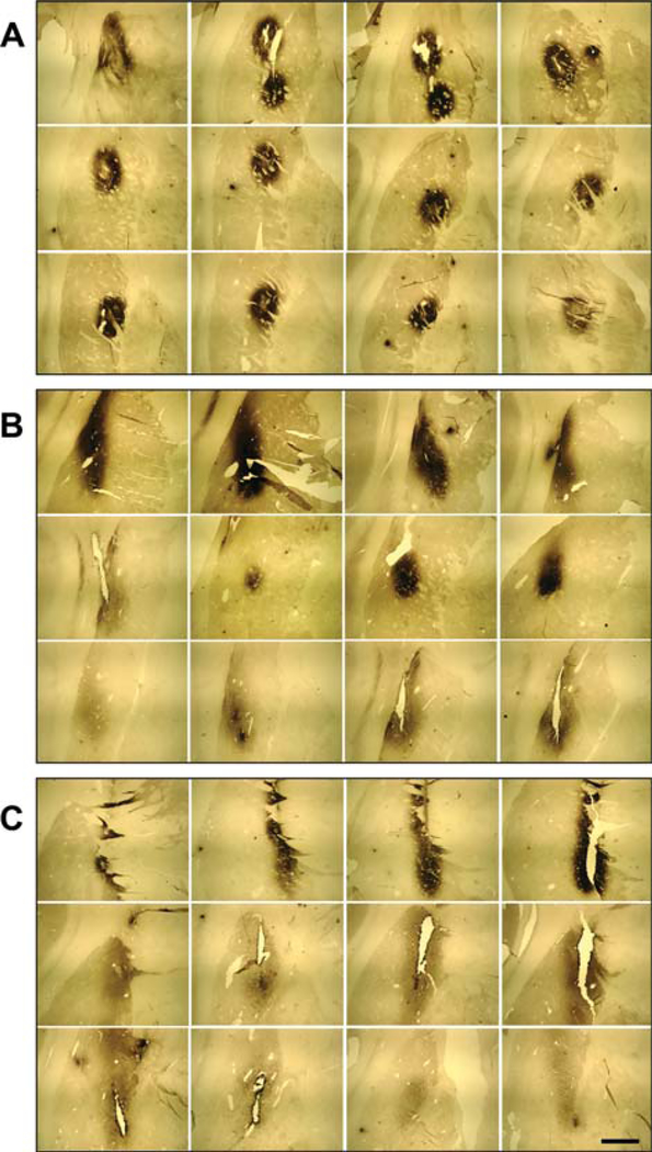
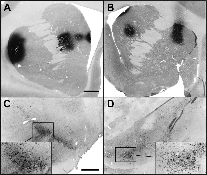
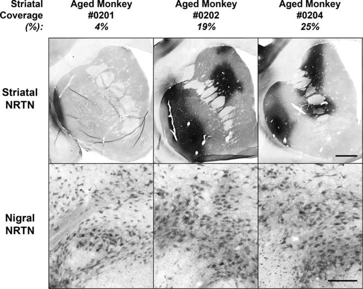
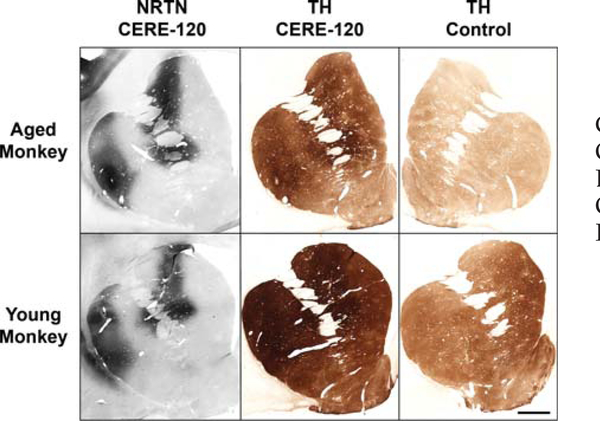
Results: NRTN-immunostaining in the targeted striatum was seen in all PD cases (mean putaminal coverage: ∼15% by volume); comparable expression was observed in young, aged, and parkinsonian monkeys. In the SNc cell bodies, however, only rare evidence of neurturin was seen in PD, while ample evidence of intense nigral-NRTN was observed in all monkeys. NRTN-expression was associated with occasional, sparse TH-induction in the striatum of PD, but nothing apparent in the SNc. In primates, NRTN produced robust TH-induction throughout the nigrostriatal neurons.

Discussion: These data provide the first evidence that gene therapy can increase expression of a neurotrophic-factor deep in the PD brain and that clear but modest enhancement of degenerating neurons can be induced. They also provide important insight regarding deficiencies in the status of nigrostriatal neurons in advanced PD, suggesting that serious axon-transport deficits reduced the bioactivity of AAV2-NRTN by limiting the protein exposed to the cell body. Thus, future efforts using neurotrophic-factors to treat neurodegenerative diseases will need to target both the terminal fields and the cell bodies of degenerating neurons to assure maximal benefit is achieved.


Conflict of interest statement

Potential conflict of interest: Several of the authors are employees of Ceregene Inc., (see affiliations), the company developing AAV2-NRTN (CERE-120) for PD. The remaining authors (except for Y.C.) are consultants to the company. Employees and consultants receive financial remuneration and have been awarded stock options from Ceregene.

Figures

FIG. 1.
FIG. 1.
Photomicrographs, illustrating neurturin (NRTN) immune-histochemical staining throughout sections of the PD putamen. A: Case 1904-left hemisphere; B: Case 1802-right hemisphere; C: Case 1802-left hemisphere. Scale bar represents 5 mm.
FIG. 2.
FIG. 2.
Example of melanin-positive neurons and NRTN-immunostaining in PD substantia nigra, pars compacta (SNc). While many clusters of melanin-positive, dopaminergic perikarya were observed in nigra (A: low magnification and B: high magnification), only rarely did any nigral neurons show evidence for NRTN staining (e.g., C: high magnification). Panels A and B illustrate the general failure of nigrostriatal neurons to retrogradely transport NRTN to nigral perikarya following intraputaminal delivery of AAV2-NRTN, whereas Panel C illustrates a rare example of NRTN in the nigra from successful retrograde transport. Scale bar represents 220 μm in panel A and 22 μm in panels B and C.
FIG. 3.
FIG. 3.
NRTN staining in young monkeys administered low dose of AAV2-NRTN to striatum, intentionally covering only 5.6% and 1.8% of striatal volume (panels A and B, respectively). Figures represent the coronal section with greatest area of NRTN protein for each monkey. Despite modest NRTN coverage of striatum, NRTN was clearly seen in nigra only 1 month after AAV2-NRTN administration (panels C and D). Scale bar represents 2 mm in panels A and B. Scale bar in panel C represents 1 mm in lower magnification photos in panels C and D, and in higher magnification photos in C and D, represents 400 μm.
FIG. 4.
FIG. 4.
NRTN in 3 aged monkeys treated with AAV2-NRTN administration to striatum. Note abundant NRTN-positive cells in substantia nigra in all 3 cases (bottom panels) following NRTN expression in striatum (top panels), even in case when volume of NRTN coverage was only 4%. Scale bar in top panels represents 2.0 mm, and 0.2 mm in lower panels.
FIG. 5.
FIG. 5.
NRTN in striatum and nigra of MPTP monkeys treated with AAV2-NRTN. Panels A and C: sections from a monkey with 8% NRTN coverage of striatum (lowest coverage in group). Panels B and D: monkey with ~23% coverage of putamen (highest coverage in group). Open arrow denotes edge of relatively small AAV2-NRTN nigra injection (note that NRTN-stained nigral neurons are seen several mm away, reflecting retrograde transport of NRTN from striatum). Scale bar (in B) represents 2 mm in panels A and B. Scale bar (in D) represents 1 mm in C and D lower magnification panels and 208 μm in C and D higher magnification insets.
FIG. 6.
FIG. 6.
Representative photomicrographs from PD putamen (top panels AC) and substantia nigra (bottom panels DF). Panels B and C provide example of TH induction in putamen, relative to NRTN expression in adjacent section (A), following AAV2-NRTN administration. Note TH signal: (i) was observed only well within the area of NRTN staining, (ii) was sparse, and (iii) occurred only ~50% of the time NRTN was seen. Panels D–F depict representative example of TH+ neurons and fibers in PD substantia nigra (D and E) and nigrostriatal pathway of the same hemisphere (F). Scale bar (in A) represents 1332 μm in A and B, 100 μm in C, E, and F and 833 μm in D.
FIG. 7.
FIG. 7.
Typical examples of TH induction in aged (top) and young (bottom) monkeys, showing relationship between NRTN expression in striatum (left panels) and corresponding TH induction (middle panels). For the aged monkey, the right-hand panel depicts TH-ir on his contralateral (untreated) side. For young monkey, a representative section from a control monkey injected with formulation-buffer is depicted. Note that in these (and typically all) monkey studies, the area of TH induction extends well beyond boundaries of NRTN staining. Scale bar represents 2 mm.

References

    1. Akerud P, Alberch J, Eketjall S, Wagner J, Arenas E. Differential effects of glial cell line-derived neurotrophic factor and neurturin on developing and adult substantia nigra dopaminergic neurons. J Neurochem 1999;73:70–78. - PubMed
    1. Horger BA, Nishimura MC, Armanini MP, et al. Neurturin exerts potent actions on survival and function of midbrain dopaminergic neurons. J Neurosci 1998;18:4929–4937. - PMC - PubMed
    1. Kordower JH, Emborg ME, Bloch J, et al. Neurodegeneration prevented by lentiviral vector delivery of GDNF in primate models of Parkinson’s disease. Science 2000;290:767–773. - PubMed
    1. Kordower JH, Herzog CD, Dass B, et al. Delivery of neurturin by AAV2 (CERE-120)-mediated gene transfer provides structural and functional neuroprotection and neurorestoration in MPTP-treated monkeys. Ann Neurol 2006;60:706–715. - PubMed
    1. Kordower JH, Palfi S, Chen EY, et al. Clinicopathological findings following intraventricular glial-derived neurotrophic factor treatment in a patient with Parkinson’s disease. Ann Neurol 1999;46:419–424. - PubMed

Publication types

MeSH terms