Comparative Study
. 2011 Apr 5;50(13):2456-63.
doi: 10.1021/bi101803k. Epub 2011 Mar 11.

Atomic structures suggest determinants of transmission barriers in mammalian prion disease

Affiliations
Comparative Study

Atomic structures suggest determinants of transmission barriers in mammalian prion disease

Marcin I Apostol et al. Biochemistry. .

Abstract

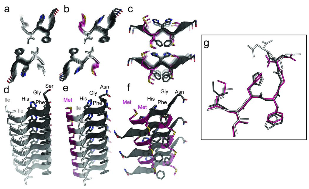
Prion represents a unique class of pathogens devoid of nucleic acid. The deadly diseases transmitted by it between members of one species and, in certain instances, to members of other species present a public health concern. Transmissibility and the barriers to transmission between species have been suggested to arise from the degree to which a pathological protein conformation from an individual of one species can seed a pathological conformation in another species. However, this hypothesis has never been illustrated at an atomic level. Here we present three X-ray atomic structures of the same segment from human, mouse, and hamster PrP, which is critical for forming amyloid and confers species specificity in PrP seeding experiments. The structures reveal that different sequences encode different steric zippers and suggest that the degree of dissimilarity of these zipper structures gives rise to transmission barriers in prion disease, such as those that protect humans from acquiring bovine spongiform encephalopathy (BSE) and chronic wasting disease (CWD).


Figures

Figure 1
Figure 1
Atomic structures from X-ray crystallography of steric zippers formed by the transmission-determining segment of human, mouse, and hamster PrP. The steric zippers of human (A), mouse (B), and hamster (C) segments 138–143 (using human numbering) are illustrated as cartoon representations showing sheet-to-sheet interactions. Side chains are drawn as stick representations, with carbon atoms in isoleucine residues highlighted in white, methionines in magenta, and the remaining residues in dark grey. In the side chains, nitrogen atoms are colored in blue, oxygen in dark red and sulfur in yellow. The bottom panels (D–F) show a view along the hydrogen bonding axis (length of the page) of one isolated sheet showing the stacking of β-strands. By comparing panels A and B it can be seen the two structures of human and mouse segments and interfaces are nearly identical. (G) The alignment of six-residue Hum138–143 (dark grey), six-reside Mus137–142 (magenta), and seven-residue Mus137–143 (white) have similar conformations with the characteristic kink at Gly142. A r.m.s.d of 0.53 Å was calculated between main-chain atoms of the human and mouse segments showing a great similarity across the aligned six residues. By contrast the r.m.s.d between the hamster/mouse and hamster/human structures is 3.427 Å and 3.169 Å, respectively.
Figure 2
Figure 2
Diagram analogizing the structural aspects of permissible strain propagation and transmission barriers with fitting a square-peg-in-a-round-hole. The left column represents template strains or seeds as a mold, the middle column represents monomers as sticks. To have successful seeding, the sticks have to fit in the mold. The right column shows the quality of the fit. The fourth column shows how this rationalization of structural fit matches the data presented by Vanik et al. and Jones et al. and summarized in Table 1 for the species dependent seeding of PrP23-144 (25, 34).
Figure 3
Figure 3
(A) The sequence alignment of the region 170–175 (using human numbering) of human (Hum), elk, hamster (Ham), bovine (Cow), mouse (Mus), sheep (Shp), and rabbit (Rab) PrP. Residues corresponding to positions 170 and 174 using human numbering have species variation, however only the elk prion has both an Asn at residue 170 and Thr at residue 174. Purple highlights the xNxNxF motif common for all species, while cyan highlights the human specific residues and yellow highlights the elk specific residues. These colors are continued in the stick representations of side chains in the human segment 170–175 (B) pdb code 3fva, and both interfaces of the homologous elk segment (C and D) pdb code 2ol9.(21, 33) The peptide backbone is depicted as a cartoon in white. In all the representations the hydrogen bonding axis of the beta-sheets is perpendicular to the plane of the page to point out the packing of side chains within the steric zipper interfaces.

References

    1. Hilton DA. Pathogenesis and prevalence of variant Creutzfeldt-Jakob disease. J. Pathol. 2006;208:134–141. - PubMed
    1. Collinge J. Variant Creutzfeldt-Jakob disease. The Lancet. 1999;354:317–323. - PubMed
    1. Aldhous P. BSE: spongiform encephalopathy found in cat. Nature. 1990;345:194. - PubMed
    1. Kirkwood JK, Cunningham AA. Epidemiological observations on spongiform encephalopathies in captive wild animals in the British Isles. Vet. Rec. 1994;135:296–303. - PubMed
    1. Sigurdson CJ, Aguzzi A. Chronic wasting disease. Biochim. Biophys. Acta. 2007;1772:610–618. - PMC - PubMed

Publication types