

PrPSc spreading patterns in the brain of sheep linked to different prion types
- PMID: 21324114
- PMCID: PMC3050706
- DOI: 10.1186/1297-9716-42-32
PrPSc spreading patterns in the brain of sheep linked to different prion types
Abstract
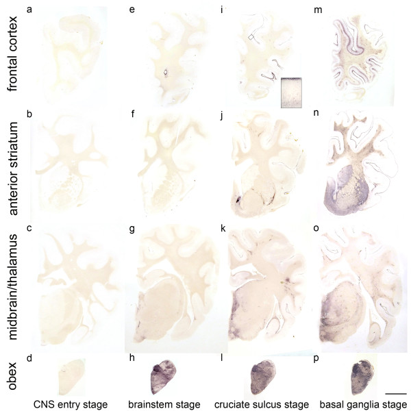
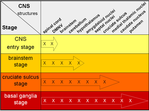
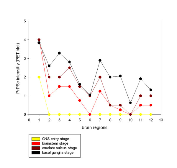
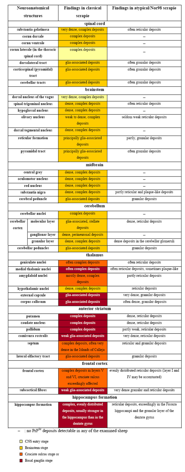
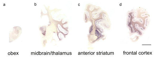
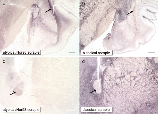
Scrapie in sheep and goats has been known for more than 250 years and belongs nowadays to the so-called prion diseases that also include e.g. bovine spongiform encephalopathy in cattle (BSE) and Creutzfeldt-Jakob disease in humans. According to the prion hypothesis, the pathological isoform (PrPSc) of the cellular prion protein (PrPc) comprises the essential, if not exclusive, component of the transmissible agent. Currently, two types of scrapie disease are known--classical and atypical/Nor98 scrapie. In the present study we examine 24 cases of classical and 25 cases of atypical/Nor98 scrapie with the sensitive PET blot method and validate the results with conventional immunohistochemistry. The sequential detection of PrPSc aggregates in the CNS of classical scrapie sheep implies that after neuroinvasion a spread from spinal cord and obex to the cerebellum, diencephalon and frontal cortex via the rostral brainstem takes place. We categorize the spread of PrPSc into four stages: the CNS entry stage, the brainstem stage, the cruciate sulcus stage and finally the basal ganglia stage. Such a sequential development of PrPSc was not detectable upon analysis of the present atypical/Nor98 scrapie cases. PrPSc distribution in one case of atypical/Nor98 scrapie in a presumably early disease phase suggests that the spread of PrPSc aggregates starts in the di- or telencephalon. In addition to the spontaneous generation of PrPSc, an uptake of the infectious agent into the brain, that bypasses the brainstem and starts its accumulation in the thalamus, needs to be taken into consideration for atypical/Nor98 scrapie.
Figures
Similar articles
-
Detection of classical and atypical/Nor98 scrapie by the paraffin-embedded tissue blot method.Vet Rec. 2009 May 30;164(22):677-81. doi: 10.1136/vr.164.22.677. Vet Rec. 2009. PMID: 19483208
-
Detection of PrP(Sc) in formalin-fixed, paraffin-embedded tissue by Western blot differentiates classical scrapie, Nor98 scrapie, and bovine spongiform encephalopathy.J Vet Diagn Invest. 2010 Sep;22(5):684-9. doi: 10.1177/104063871002200502. J Vet Diagn Invest. 2010. PMID: 20807921
-
Co-existence of classical scrapie and Nor98 in a sheep from an Italian outbreak.Res Vet Sci. 2010 Jun;88(3):478-85. doi: 10.1016/j.rvsc.2009.11.015. Epub 2009 Dec 23. Res Vet Sci. 2010. PMID: 20031179
-
The immunodetection of the abnormal isoform of prion protein.Histochem J. 1999 Apr;31(4):209-12. doi: 10.1023/a:1003514021800. Histochem J. 1999. PMID: 10447061 Review.
-
Genetic and infectious prion diseases.Arch Neurol. 1993 Nov;50(11):1129-53. doi: 10.1001/archneur.1993.00540110011002. Arch Neurol. 1993. PMID: 8105771 Review.
Cited by
-
Genome-Wide Methylation Profiling in the Thalamus of Scrapie Sheep.Front Vet Sci. 2022 Feb 16;9:824677. doi: 10.3389/fvets.2022.824677. eCollection 2022. Front Vet Sci. 2022. PMID: 35252421 Free PMC article.
-
Cerebellar compartmentation of prion pathogenesis.Brain Pathol. 2018 Mar;28(2):240-263. doi: 10.1111/bpa.12503. Epub 2017 Apr 10. Brain Pathol. 2018. PMID: 28268246 Free PMC article.
-
Evolutionary biology and the risk of scrapie disease in sheep.Open Vet J. 2018;8(3):282-294. doi: 10.4314/ovj.v8i3.7. Epub 2018 Aug 7. Open Vet J. 2018. PMID: 30148080 Free PMC article. Review.
-
Atypical scrapie isolates involve a uniform prion species with a complex molecular signature.PLoS One. 2011;6(11):e27510. doi: 10.1371/journal.pone.0027510. Epub 2011 Nov 11. PLoS One. 2011. PMID: 22096587 Free PMC article.
-
The interpretation of disease phenotypes to identify TSE strains following murine bioassay: characterisation of classical scrapie.Vet Res. 2012 Nov 1;43(1):77. doi: 10.1186/1297-9716-43-77. Vet Res. 2012. PMID: 23116457 Free PMC article.
References
-
- McGowan JP. Scrapie in sheep. Scottish J Agric. 1922;5:365–375.
-
- Bendheim PE, Brown HR, Rudelli RD, Scala LJ, Goller NL, Wen GY, Kascsak RJ, Cashman NR, Bolton DC. Nearly ubiquitous tissue distribution of the scrapie agent precursor protein. Neurology. 1992;42:149–156. - PubMed
Publication types
MeSH terms
Substances
LinkOut - more resources
Full Text Sources
