

SDF1 in the dorsal corticospinal tract promotes CXCR4+ cell migration after spinal cord injury
- PMID: 21324162
- PMCID: PMC3050722
- DOI: 10.1186/1742-2094-8-16
SDF1 in the dorsal corticospinal tract promotes CXCR4+ cell migration after spinal cord injury
Erratum in
- J Neuroinflammation. 2011;8:37. Belmadani, Abdelhak [added]
-
Erratum to: SDF1 in the dorsal corticospinal tract promotes CXCR4+ cell migration after spinal cord injury.J Neuroinflammation. 2017 Feb 14;14(1):35. doi: 10.1186/s12974-017-0810-0. J Neuroinflammation. 2017. PMID: 28196519 Free PMC article. No abstract available.
Abstract
Background: Stromal cell-derived factor-1 (SDF1) and its major signaling receptor, CXCR4, were initially described in the immune system; however, they are also expressed in the nervous system, including the spinal cord. After spinal cord injury, the blood brain barrier is compromised, opening the way for chemokine signaling between these two systems. These experiments clarified prior contradictory findings on normal expression of SDF1 and CXCR4 as well as examined the resulting spinal cord responses resulting from this signaling.
Methods: These experiments examined the expression and function of SDF1 and CXCR4 in the normal and injured adult mouse spinal cord primarily using CXCR4-EGFP and SDF1-EGFP transgenic reporter mice.
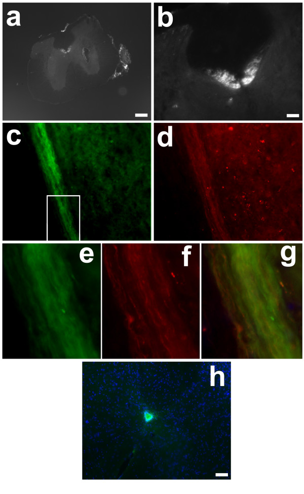
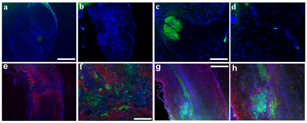
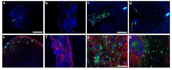
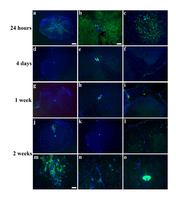
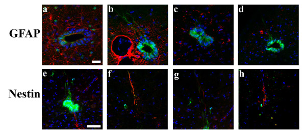
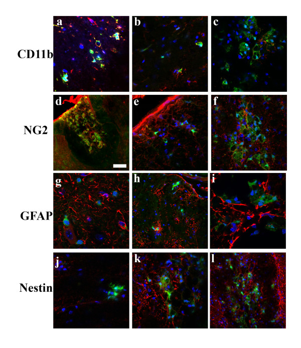
Results: In the uninjured spinal cord, SDF1 was expressed in the dorsal corticospinal tract (dCST) as well as the meninges, whereas CXCR4 was found only in ependymal cells surrounding the central canal. After spinal cord injury (SCI), the pattern of SDF1 expression did not change rostral to the lesion but it disappeared from the degenerating dCST caudally. By contrast, CXCR4 expression changed dramatically after SCI. In addition to the CXCR4+ cells in the ependymal layer, numerous CXCR4+ cells appeared in the peripheral white matter and in the dorsal white matter localized between the dorsal corticospinal tract and the gray matter rostral to the lesion site. The non-ependymal CXCR4+ cells were found to be NG2+ and CD11b+ macrophages that presumably infiltrated through the broken blood-brain barrier. One population of macrophages appeared to be migrating towards the dCST that contains SDF1 rostral to the injury but not towards the caudal dCST in which SDF1 is no longer present. A second population of the CXCR4+ macrophages was present near the SDF1-expressing meningeal cells.
Conclusions: These observations suggest that attraction of CXCR4+ macrophages is part of a programmed response to injury and that modulation of the SDF1 signaling system may be important for regulating the inflammatory response after SCI.
Figures
Similar articles
-
Concise review: the potential of stromal cell-derived factor 1 and its receptors to promote stem cell functions in spinal cord repair.Stem Cells Transl Med. 2012 Oct;1(10):732-9. doi: 10.5966/sctm.2012-0068. Epub 2012 Oct 10. Stem Cells Transl Med. 2012. PMID: 23197665 Free PMC article. Review.
-
Effect of SDF-1/CXCR4 axis on the migration of transplanted bone mesenchymal stem cells mobilized by erythropoietin toward lesion sites following spinal cord injury.Int J Mol Med. 2015 Nov;36(5):1205-14. doi: 10.3892/ijmm.2015.2344. Epub 2015 Sep 14. Int J Mol Med. 2015. PMID: 26398409 Free PMC article.
-
Biphasic MIF and SDF1 expression during podocyte injury promote CD44-mediated glomerular parietal cell migration in focal segmental glomerulosclerosis.Am J Physiol Renal Physiol. 2020 Mar 1;318(3):F741-F753. doi: 10.1152/ajprenal.00414.2019. Epub 2020 Feb 18. Am J Physiol Renal Physiol. 2020. PMID: 32068458
-
SDF1/CXCR4 signalling regulates two distinct processes of precerebellar neuronal migration and its depletion leads to abnormal pontine nuclei formation.Development. 2009 Jun;136(11):1919-28. doi: 10.1242/dev.032276. Development. 2009. PMID: 19429788
-
CXCL12 signaling in the development of the nervous system.J Neuroimmune Pharmacol. 2012 Dec;7(4):820-34. doi: 10.1007/s11481-011-9336-x. Epub 2012 Jan 21. J Neuroimmune Pharmacol. 2012. PMID: 22270883 Free PMC article. Review.
Cited by
-
Achieving CNS axon regeneration by manipulating convergent neuro-immune signaling.Cell Tissue Res. 2012 Jul;349(1):201-13. doi: 10.1007/s00441-012-1425-5. Epub 2012 May 17. Cell Tissue Res. 2012. PMID: 22592625 Free PMC article. Review.
-
Spinal cord injury and inflammatory mediators: Role in "fire barrier" formation and potential for neural regeneration.Neural Regen Res. 2026 Mar 1;21(3):923-937. doi: 10.4103/NRR.NRR-D-24-00792. Epub 2025 Feb 24. Neural Regen Res. 2026. PMID: 39995083 Free PMC article.
-
Glial cells activation potentially contributes to the upregulation of stromal cell-derived factor-1α after optic nerve crush in rats.Neurochem Res. 2013 Oct;38(10):1996-2008. doi: 10.1007/s11064-013-1106-0. Epub 2013 Jul 6. Neurochem Res. 2013. PMID: 23832528
-
Regulation of inflammatory cytokines for spinal cord injury recovery.Histol Histopathol. 2021 Feb;36(2):137-142. doi: 10.14670/HH-18-262. Epub 2020 Oct 1. Histol Histopathol. 2021. PMID: 33001420 Review.
-
Concise review: the potential of stromal cell-derived factor 1 and its receptors to promote stem cell functions in spinal cord repair.Stem Cells Transl Med. 2012 Oct;1(10):732-9. doi: 10.5966/sctm.2012-0068. Epub 2012 Oct 10. Stem Cells Transl Med. 2012. PMID: 23197665 Free PMC article. Review.
References
-
- Klein RS, Rubin JB, Gibson HD, DeHaan EN, Alvarez-Hernandez X, Segal RA, Luster AD. SDF-1 alpha induces chemotaxis and enhances Sonic hedgehog-induced proliferation of cerebellar granule cells. Development. 2001;128:1971–1981. - PubMed
Publication types
MeSH terms
Substances
Grants and funding
LinkOut - more resources
Full Text Sources
Other Literature Sources
Medical
Molecular Biology Databases
Research Materials
