. 2011 May;32(14):3575-83.
doi: 10.1016/j.biomaterials.2011.01.062. Epub 2011 Feb 13.

The role of extracellular matrix composition in structure and function of bioengineered skeletal muscle

Affiliations

The role of extracellular matrix composition in structure and function of bioengineered skeletal muscle

Sara Hinds et al. Biomaterials. 2011 May.

Abstract

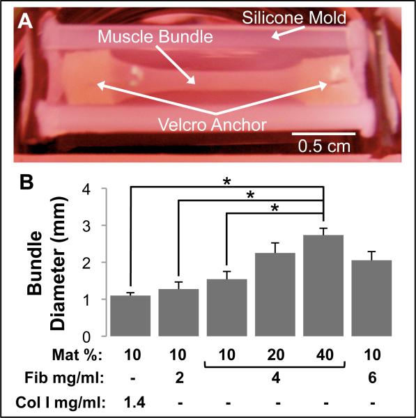
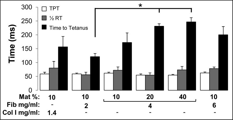
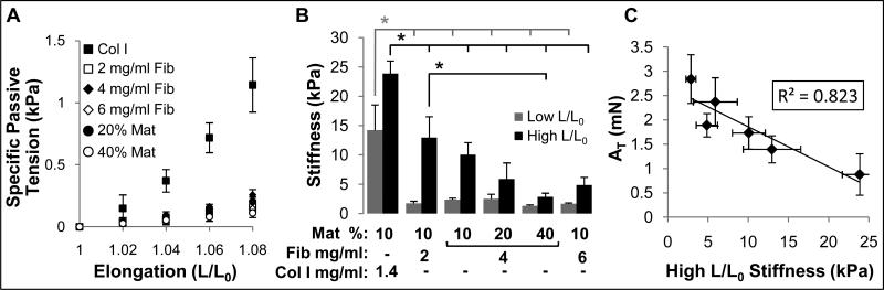
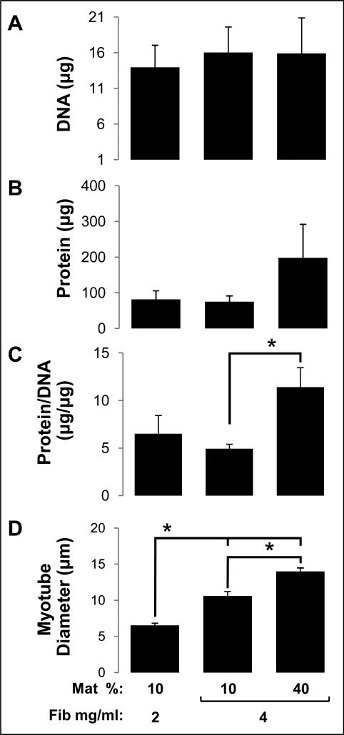
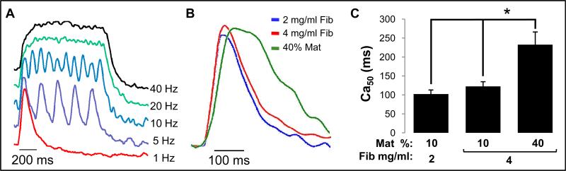
One of the obstacles to the potential clinical utility of bioengineered skeletal muscle is its limited force generation capacity. Since engineered muscle, unlike most native muscle tissue, is composed of relatively short myofibers, we hypothesized that, its force production and transmission would be profoundly influenced by cell-matrix interactions. To test this hypothesis, we systematically varied the matrix protein type (collagen I/fibrin/Matrigel) and concentration in engineered, hydrogel-based neonatal rat skeletal muscle bundles and assessed the resulting tissue structure, generation of contractile force, and intracellular Ca(2+) handling. After two weeks of culture, the muscle bundles consisted of highly aligned and cross-striated myofibers and exhibited standard force-length and force-frequency relationships achieving tetanus at 40 Hz. The use of 2 mg/ml fibrin (control) yielded isometric tetanus amplitude of 1.4 ± 0.3 mN as compared to 0.9 ± 0.4 mN measured in collagen I-based bundles. Higher fibrin and Matrigel concentrations synergistically yielded further increase in active force generation to 2.8 ± 0.5 mN without significantly affecting passive mechanical properties, tetanus-to-twitch ratio, and twitch kinetics. Optimized matrix composition yielded significant cellular hypertrophy (protein/DNA ratio = 11.4 ± 4.1 vs. 6.5 ± 1.9 μg/μg in control) and a prolonged Ca(2+) transient half-width (Ca(50) = 232.8 ± 33.3 vs. 101.7 ± 19.8 ms). The use of growth-factor-reduced Matrigel, instead of standard Matrigel did not alter the obtained results suggesting enhanced cell-matrix interactions rather than growth factor supplementation as an underlying cause for the measured increase in contractile force. In summary, biomaterial-based manipulation of cell-matrix interactions represents an important target for improving contractile force generation in engineered skeletal muscle.


Figures

Figure 1
Figure 1
Morphology of bioengineered skeletal muscle bundles. (A) A representative muscle bundle shown in a silicone tissue mold. The muscle bundle was anchored at each end to pinned Velcro® tabs. (B) Final muscle bundle diameter on culture day 14 for the 6 studied hydrogel formulations. The hydrogel formulations contained different volume percent Matrigel (Mat %), fibrinogen concentration (Fib mg/ml) and collagen I concentration (Col I mg/ml). Bars show n=3 bundles per group. ‘*’, statistically significant between the indicated pairings, p<0.05.
Figure 2
Figure 2
Structure of bioengineered skeletal muscle bundles. (A1-3) Representative H&E stainings of a day 14 muscle bundle showing densely packed myotubes, located in the outer bundle region (A1), that were longitudinally oriented (A2-3). White arrow in A3 indicates visible cross-striations. (B) Confocal stack image of a day 14 bundle. Top and right frames are front and side views of the stack, respectively. Note that fibroblasts (red) form a single outermost layer on the bundle surface and cover muscle layers underneath (green). (C) A representative confocal section ~20 μm below the bundle surface shows that aligned multinucleated myotubes exhibit ubiquitous cross-striations. (D) Typical immunostaining for α7 integrins (green, white arrows) along myotube membrane. Panels A1-3, B, and D are from muscle bundles with 4 mg/ml fibrinogen and 10% Matrigel, while panel C is from a muscle bundle with 4 mg/ml fibrinogen and 40% Matrigel.
Figure 3
Figure 3
Contractile force generation in bioengineered skeletal muscle bundles. (A,B) Peak twitch (At) and tetanus (AT) amplitudes for the 6 studied hydrogel formulations. The nomenclature describing hydrogel formulations is the same as shown in figure 1. Bars from left to right are obtained from n=4, 7, 8, 7, 7, and 6 bundles per group. ‘*’, statistically significant between the indicated pairings, p<0.05.
Figure 4
Figure 4
Kinetics of contractile force generation in bioengineered skeletal muscle bundles. Time to peak twitch (TPT), half-relaxation time (1/2RT), and time-to-tetanus are shown for the 6 studied hydrogel formulations. The nomenclature describing hydrogel formulations and number of bundles per group are the same as shown in figure 3. ‘*’, statistically significant between the indicated pairings, p<0.05.
Figure 5
Figure 5
Passive mechanical properties of bioengineered skeletal muscle bundles. (A) Specific passive tension (stress) as a function of bundle elongation (L/L0) shown for the 6 studied hydrogel formulations. Note increased stress in collagen I-based vs. fibrin-based bundles. The groups without indicated Mat% contain 10% Matrigel, while those with only indicated % Mat contain 4 mg/ml fibrinogen. (B) Muscle bundle stiffness at high and low L/L0. The nomenclature describing hydrogel formulations (bundle groups) is the same as shown in figure 1. (C) Correlation between tetanus amplitude (AT) and bundle stiffness at high elongation in all 6 studied bundle groups. Number of bundles per group is the same as in figure 3. ‘*’, statistically significant between the indicated pairings, p<0.05.
Figure 6
Figure 6
Indices of cell number and size in fibrin-based skeletal muscle bundles. (A) Total DNA content (index of cell number) in bundles. (B) Total cellular protein content in bundles. (C) Cellular protein/DNA ratio (index of average cell size) in bundles. (D) Average myotube diameter in bundles. The nomenclature describing hydrogel formulations is the same as in figure 1. For A-C, n=4 bundles per group; For D, n=3 bundles per group. ‘*’, statistically significant between the indicated pairings, p<0.05.
Figure 7
Figure 7
Intracellular Ca2+ transients in fibrin-based skeletal muscle bundles. (A) Representative optically recorded traces of intracellular Ca2+ concentration for different stimulation frequencies. Note that increasing pacing frequency yields a sustained elevated level of intracellular Ca2+ concentration. (B) Representative single Ca2+ transients for 3 studied hydrogel compositions showing differences in the transient duration. (C) Half-width of the Ca2+ transient duration (Ca50) in engineered bundles. Bars show n=3 bundles per group. The nomenclatures describing hydrogel compositions are the same as in figure 5. ‘*’, statistically significant between the indicated pairings, p<0.05.
Figure 8
Figure 8
Comparison of contractile force generation in bioengineered skeletal muscle bundles made using standard vs. reduced growth factor (RGF) Matrigel. (A) Twitch (At) and tetanus (AT) amplitudes. (B) Kinetic parameters of twitch and tetanus, with nomenclature defined in figure 4. Muscle bundles were made with 4 mg/ml Fibrinogen and 40% standard or RGF Matrigel. Bars show n=3 bundles per group.

References

    1. Alsberg E, Hill EE, Mooney DJ. Craniofacial tissue engineering. Crit Rev Oral Biol Med. 2001;12:64–75. - PubMed
    1. Bach AD, Beier JP, Stern-Staeter J, Horch RE. Skeletal muscle tissue engineering. J Cell Mol Med. 2004;8:413–22. - PMC - PubMed
    1. Vilquin JT. Myoblast transplantation: clinical trials and perspectives. Mini-review. Acta Myol. 2005;24:119–27. - PubMed
    1. Bian W, Bursac N. Tissue engineering of functional skeletal muscle: challenges and recent advances. IEEE Eng Med Biol Mag. 2008;27:109–13. - PMC - PubMed
    1. Liao H, Zhou GQ. Development and progress of engineering of skeletal muscle tissue. Tissue Eng Part B Rev. 2009;15:319–31. - PubMed

Publication types