. 2011 Apr 22;286(16):14324-34.
doi: 10.1074/jbc.M111.218164. Epub 2011 Feb 15.

Subunit dimers of alpha-hemolysin expand the engineering toolbox for protein nanopores

Affiliations

Subunit dimers of alpha-hemolysin expand the engineering toolbox for protein nanopores

Anne F Hammerstein et al. J Biol Chem. .

Abstract

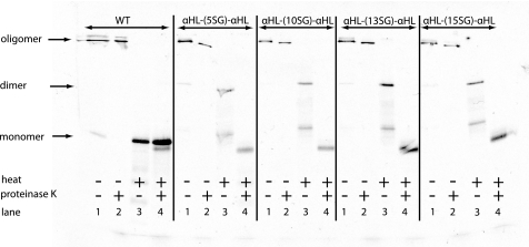
Staphylococcal α-hemolysin (αHL) forms a heptameric pore that features a 14-stranded transmembrane β-barrel. We attempted to force the αHL pore to adopt novel stoichiometries by oligomerizing subunit dimers generated by in vitro transcription and translation of a tandem gene. However, in vitro transcription and translation also produced truncated proteins, monomers, that were preferentially incorporated into oligomers. These oligomers were shown to be functional heptamers by single-channel recording and had a similar mobility to wild-type heptamers in SDS-polyacrylamide gels. Purified full-length subunit dimers were then prepared by using His-tagged protein. Again, single-channel recording showed that oligomers made from these dimers are functional heptamers, implying that one or more subunits are excluded from the central pore. Therefore, the αHL pore resists all structures except those that possess seven subunits immediately surrounding the central axis. Although we were not able to change the stoichiometry of the central pore of αHL by the concatenation of subunits, we extended our findings to prepare pores containing one subunit dimer and five monomers and purified them by SDS-PAGE. Two half-chelating ligands were then installed at adjacent sites, one on each subunit of the dimer. Single-channel recording showed that pores formed from this construct formed complexes with divalent metal ions in a similar fashion to pores containing two half-chelating ligands on the same subunit, confirming that the oligomers had assembled with seven subunits around the central lumen. The ability to incorporate subunit dimers into αHL pores increases the range of structures that can be obtained from engineered protein nanopores.


Figures

FIGURE 1.
FIGURE 1.
Separation of heteroheptamers generated from αHL and αHL-D8 and possible stoichiometries of pores containing subunit dimers of αHL. A, heteroheptamers generated from the monomeric subunits αHL and/or αHL-D8 (figure adapted from Ref. 42). B, WT αHL forms heptameric pores with seven subunits arranged around a central axis (20177).4 Subunit dimers of αHL could adopt novel stoichiometries, such as an octamer comprising four subunit dimers (24108) or a hexamer comprising three subunit dimers (23106).
FIGURE 2.
FIGURE 2.
Structural considerations for the design of subunit dimers of αHL. A, possible arrangements of subunit dimers of αHL within oligomers. (1) An octamer comprising four subunit dimers arranged to form the central pore (24108).4 (2) The subunits from the subunit dimers are interspersed, in non-adjacent positions in the oligomer. (3) A subunit dimer spans two oligomers, both of which are 24107 oligomers. (4) A subunit is excluded from the central pore of the oligomer (24107). B, the structure of the αHL heptamer (PDB code 7AHL) was used to determine the length of the linker required to connect the C terminus of the first subunit (red) in a subunit dimer to the N terminus of the second subunit (yellow). In a PyMOL model, we estimated the distance across the protein surface from the C terminus (blue) via Phe-42 (cyan) and Thr-12 (purple) (all on the red first subunit) to the N terminus (green) on the yellow second subunit to be 37 Å. This represents the minimum length of the linker if the N terminus of the second (yellow) subunit is not displaced.
FIGURE 3.
FIGURE 3.
Design of the serine-glycine peptide linkers. A, schematic of the subunit dimer gene in a pT7 vector. The 3′ end of one full-length gene of αHL (red) was connected to the 5′ end of a second full-length gene (yellow) through a peptide linker (blue). B, DNA and amino acid sequences of linkers containing from 5 to 15 serines and glycines. C, the lengths of the linkers in B were estimated from PyMOL models of the corresponding peptide sequences by measuring the through-space distance between the Cα atoms at the termini of the elongated peptides.
FIGURE 4.
FIGURE 4.
Expression and hemolytic activity of subunit dimers of αHL. A, 10% BisTris polyacrylamide gel showing monomers and dimers of αHL synthesized by coupled IVTT. Lane 1, WT αHL monomer; lane 2, αHL-(5SG)-αHL subunit dimer; lane 3, αHL-(10SG)-αHL dimer; lane 4, αHL-(13SG)-αHL dimer; lane 5, αHL-(15SG)-αHL dimer. B, 5% Tris·HCl SDS-polyacrylamide gel showing oligomers formed by monomers and dimers of αHL on rRBCMs. The proteins in lanes 1–5 are the same as those shown in A. The two closely spaced diffuse bands of oligomers formed by the subunit dimer preparations are indicated by arrows. C, hemolytic activity assay with rabbit erythrocytes. Monomers and dimers of αHL were synthesized by IVTT and serially 2-fold diluted across the row of a microtiter plate. Pore formation was initiated by adding washed rabbit erythrocytes, and the rate of hemolysis was monitored by observing the decrease in light scattering at 595 nm over 1 h.
FIGURE 5.
FIGURE 5.
Limited proteolysis assay to probe the conformation of the N termini and linkers in oligomers formed from subunit dimers of αHL. SDS-PAGE-purified oligomers of αHL in 20 mm Tris·HCl buffer, pH 8.0, were split into four equal samples and subjected to different treatments. Lane 1, no treatment; lane 2, proteinase K (0.5 mg/ml final) for 5 min, sample not heated; lane 3, no proteinase K, sample heated to 95 °C for 10 min in SDS-containing sample buffer; lane 4, proteinase K (0.5 mg/ml final) for 5 min and heated to 95 °C for 10 min in SDS-containing sample buffer.
FIGURE 6.
FIGURE 6.
Oligomers made from His-tagged subunit dimers of αHL that had been purified by Ni-NTA affinity chromatography. A, monomers and dimers of αHL in a 12% BisTris polyacrylamide gel. Lane 1, WT αHL monomer, no purification; lane 2, αHL-H6 monomer, purified with Ni-NTA magnetic beads; lane 3, αHL-(10SG)-αHL subunit dimer, no purification; lane 4, αHL-(10SG)-αHL-H6 subunit dimer, purified with Ni-NTA magnetic beads. B, oligomers of αHL in a 5% Tris·HCl SDS-polyacrylamide gel. Lane 1, oligomers from WT αHL monomer; lane 2, oligomers from αHL-(10SG)-αHL dimer, no purification; lane 3, oligomers from αHL-(10SG)-αHL-H6 subunit dimers, purified with Ni-NTA magnetic beads. C, oligomer of purified αHL-(10SG)-αHL-H6, cut from a 5% Tris·HCl SDS-polyacrylamide gel (B), heated to 95 °C for 10 min in SDS-containing sample buffer and re-run in a 12% BisTris polyacrylamide gel.
FIGURE 7.
FIGURE 7.
Representative pore insertion events and unitary conductance histograms. αHL oligomers were extracted from SDS-polyacrylamide gels, before insertion into planar lipid bilayers. The oligomers were made from A, WT αHL. B, subunit dimers of αHL. C, Ni-NTA-purified subunit dimers of αHL. D, Ni-NTA-purified subunit dimers of αHL, and the oligomers were treated with proteinase K before the recording was made. The current traces were recorded in 2.0 m KCl, 10 mm MOPS, pH 7.0, at an applied potential of −50 mV.
FIGURE 8.
FIGURE 8.
Comparison of oligomers made from monomers of αHL and Ni-NTA-purified subunit dimers of αHL. Oligomers made from subunit dimers were treated with proteinase K. A, current-voltage (I-V) relationships obtained in 2.0 m KCl, 10 mm MOPS, pH 7.0. Mean values ± S.D. for each point were calculated from at least five different pores. B, single channel recording traces showing β-cyclodextrin binding to oligomers of αHL. The recordings were obtained in 2.0 m KCl, 10 mm MOPS, pH 7.0, at −50 mV. β-Cyclodextrin (40 μm) was added to the trans recording chamber. The mean residence time (τoff) of βCD in the pore was 0.39 ± 0.03 ms (n = 5) for WT pores and 0.33 ± 0.03 ms (n = 5) for oligomers made from subunit dimers. The inter-event intervals (τon) were 84 ± 14 and 115 ± 14 ms, respectively.
FIGURE 9.
FIGURE 9.
Composition of oligomers containing monomers and subunit dimers of αHL. A, mixtures of monomers and dimers of αHL were assembled on rRBCM and subjected to electrophoresis in a 5% SDS-polyacrylamide gel. The monomers carried a D8H6 tail to allow separation of the oligomers. Lane A, dimer/monomer ratio 5:0; lane B, dimer/monomer 4:1; lane C, dimer/monomer 3:2; lane D, dimer/monomer 2:3; lane E, dimer/monomer 1:4; lane F, dimer/monomer 0:5. B, the experiment in A was repeated on a larger scale, and the samples with different ratios of monomer to dimer were combined (“Mix”). C, the oligomers in bands 1–4 from B were excised from the gel and heated to 95 °C for 10 min to disrupt the subunit interactions. The polypeptides were then subjected to electrophoresis in a 12% SDS-polyacrylamide gel to resolve the constituents, and the relative intensities of the bands were measured from an autoradiogram by using Quantity One software (Bio-Rad). The predicted intensities of the bands is given in parentheses for the following are: lane 1, 20177; lane 2, 21157; lane 3, 22137; lane 4, 23117. D, proposed stoichiometries of the oligomers 1–4 in B.
FIGURE 10.
FIGURE 10.
Half-chelators installed on two adjacent subunits in the αHL heptamer. A, structure of the half-chelator reagent containing the PIDA group. B, molecular model of the P(pida)2′ pore. The two half-chelators are attached to cysteine residues at position 145 on the first subunit (red) and position 117 on the second subunit (yellow) of the subunit dimer. C, single channel recordings at −50 mV with a buffer containing 2.0 m KCl, 2 mm succinic acid, pH 4.0, in both chambers and 500 μm ZnCl2 in the trans chamber. The Ppida′ pore contains a single half-chelator at position 117, while the P(pida)2′ pore contains two half-chelators, one at each of positions 145 and 117. The four current levels for the P(pida)2′ pore correspond to the unoccupied pore, a single Zn2+ ion bound to one or the other of the half-chelators (levels A and B), and Zn2+ fully complexed with both half-chelators (level C). D, proposed kinetic scheme.

Similar articles

Cited by

References

    1. Howorka S., Siwy Z. (2009) Chem. Soc. Rev. 38, 2360–2384 - PubMed
    1. Majd S., Yusko E. C., Billeh Y. N., Macrae M. X., Yang J., Mayer M. (2010) Curr. Opin. Biotechnol. 21, 439–476 - PMC - PubMed
    1. Siwy Z. S., Howorka S. (2010) Chem. Soc. Rev. 39, 1115–1132 - PubMed
    1. Panchal R. G., Cusack E., Cheley S., Bayley H. (1996) Nat. Biotechnol. 14, 852–856 - PubMed
    1. Eroglu A., Russo M. J., Bieganski R., Fowler A., Cheley S., Bayley H., Toner M. (2000) Nat. Biotechnol. 18, 163–167 - PubMed

Publication types

LinkOut - more resources