. 2011 Mar;4(2):165-78.
doi: 10.1242/dmm.004077. Epub 2011 Feb 14.

Remodeling and homeostasis of the extracellular matrix: implications for fibrotic diseases and cancer

Affiliations

Remodeling and homeostasis of the extracellular matrix: implications for fibrotic diseases and cancer

Thomas R Cox et al. Dis Model Mech. 2011 Mar.

Abstract

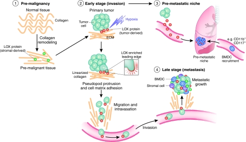
Dynamic remodeling of the extracellular matrix (ECM) is essential for development, wound healing and normal organ homeostasis. Life-threatening pathological conditions arise when ECM remodeling becomes excessive or uncontrolled. In this Perspective, we focus on how ECM remodeling contributes to fibrotic diseases and cancer, which both present challenging obstacles with respect to clinical treatment, to illustrate the importance and complexity of cell-ECM interactions in the pathogenesis of these conditions. Fibrotic diseases, which include pulmonary fibrosis, systemic sclerosis, liver cirrhosis and cardiovascular disease, account for over 45% of deaths in the developed world. ECM remodeling is also crucial for tumor malignancy and metastatic progression, which ultimately cause over 90% of deaths from cancer. Here, we discuss current methodologies and models for understanding and quantifying the impact of environmental cues provided by the ECM on disease progression, and how improving our understanding of ECM remodeling in these pathological conditions is crucial for uncovering novel therapeutic targets and treatment strategies. This can only be achieved through the use of appropriate in vitro and in vivo models to mimic disease, and with technologies that enable accurate monitoring, imaging and quantification of the ECM.


Figures

Fig. 1.
Fig. 1.
Variations in tissue stiffness. The biomechanical properties of a tissue in terms of stiffness (elastic modulus), measured in pascals (Pa), vary markedly between organs and tissues, and are inherently related to tissue function. Mechanically static tissues such as brain or compliant tissues such as lung exhibit low stiffness, whereas tissues exposed to high mechanical loading, such as bone or skeletal muscle, exhibit elastic moduli with a stiffness that is several orders of magnitude greater. Tumorigenesis is typically associated with an increase in matrix and tissue stiffness, as in breast cancer. Adapted, with permission, from Butcher et al. (Butcher et al., 2009).
Fig. 2.
Fig. 2.
Modeling mammary epithelial cells in vitro. Schematic to show modeling of mammary epithelial cells (MECs) in 3D assays in vitro. MECs form organized and polarized acini structures when grown in reconstituted basement membrane in vitro. Milk production can be induced through stimulation with lactogenic hormones. A transformation event results in cell invasion into the lumen. Increased invasive ability correlates with the development of disorganized and branching structures. Increasing matrix stiffness can also induce these events. Adapted, with permission, from Kass et al. (Kass et al., 2007) and Butcher et al. (Butcher et al., 2009).
Fig. 3.
Fig. 3.
Second harmonic generation (SHG) imaging of collagen fibril linearization during mammary gland tumorigenesis. Images are representative of whole, unfixed mammary glands of MMTV-neu mice [which carry an activated neu oncogene driven by a mouse mammary tumor virus (MMTV) promoter] and show that collagen fibril linearity increases with malignant progression, correlating with increased tissue stiffness. Arrowheads indicate linearized collagen fibrils. Image adapted, with permission, from Levental et al. (Levental et al., 2009).
Fig. 4.
Fig. 4.
Imaging the biomechanical properties of the matrix. Breast tumors are typically identified by changes in tissue mechanics, which can be detected physically, through palpitation or via imaging modalities that exploit tumor-associated changes. (A,B) Images of human breast tumor identified by ultrasound echogram (A) and mammary elastography imaging (B). The dashed lines roughly outline the imaged lesion boundary. The elastogram seems to shows a larger apparent lesion width but a similar height, relative to the echogram. This seems to be due to lateral protrusions, which are consistent with a desmoplastic response associated with local invasion that takes advantage of existing ductal and vascular anatomy. (C,D) MRI images of a 1-methyl-1-nitrosourea (MNU)-induced mammary carcinoma in rat. Here, not only do the changes in tumor ECM provide enhanced tissue contrast, but the intrinsic susceptibility of MRI exploits the paramagnetic properties of deoxyhemoglobin in erythrocytes. Deoxyhemoglobin therefore acts as an intrinsic, blood-oxygenation-level-dependent contrast agent, further highlighting the highly vascular nature of tumors. Ultrasound and elastography images were supplied courtesy of Jeff Bamber (The Institute of Cancer Research, UK). MRI images were supplied courtesy of Simon P. Robinson (The Institute of Cancer Research, UK).
Fig. 5.
Fig. 5.
The role of LOX in tumor progression. Schematic to summarize the role of LOX in tumor progression. (1) Increased LOX expression from stromal cells results in increased collagen linearization and tissue stiffness in pre-malignant tissue. These changes increase tumor incidence and tumor burden, and drive malignant progression. (2) LOX secreted by hypoxic tumor cells increases invasion, enabling metastatic dissemination. (3) LOX secreted by hypoxic tumor cells accumulates at distant sites of future metastasis and recruits BMDCs to form the pre-metastatic niche. This greatly enables establishment and growth of metastases. (4) LOX-expressing tumor cells have an enhanced ability to colonize distant organs and form metastases. LOX secreted by both tumor and stromal cells supports metastatic tumor growth. Adapted, with permission, from Erler and Giaccia (Erler and Giaccia, 2008).

References

    1. Abraham D. J., Varga J. (2005). Scleroderma: from cell and molecular mechanisms to disease models. Trends Immunol. 26, 587–595 - PubMed
    1. Aggarwal B. B., Gehlot P. (2009). Inflammation and cancer: how friendly is the relationship for cancer patients? Curr. Opin. Pharmacol. 9, 351–369 - PMC - PubMed
    1. Ahn G. O., Brown J. M. (2008). Matrix metalloproteinase-9 is required for tumor vasculogenesis but not for angiogenesis: role of bone marrow-derived myelomonocytic cells. Cancer Cell 13, 193–205 - PMC - PubMed
    1. Akhtar R., Sherratt M. J., Watson R. E., Kundu T., Derby B. (2009a). Mapping the micromechanical properties of cryo-sectioned aortic tissue with scanning acoustic microscopy. Mater. Res. Soc. Symp. Proc. 1132E - PMC - PubMed
    1. Akhtar R., Schwarzer N., Sherratt M. J., Watson R. E., Graham H. K., Trafford A. W., Mummery P. M., Derby B. (2009b). Nanoindentation of histological specimens: mapping the elastic properties of soft tissues. J. Mater. Res. 24, 638–646 - PMC - PubMed

Publication types