. 2011 Mar 15;124(Pt 6):879-91.
doi: 10.1242/jcs.076976. Epub 2011 Feb 15.

Kindlin-2 regulates podocyte adhesion and fibronectin matrix deposition through interactions with phosphoinositides and integrins

Affiliations

Kindlin-2 regulates podocyte adhesion and fibronectin matrix deposition through interactions with phosphoinositides and integrins

Hong Qu et al. J Cell Sci. .

Abstract

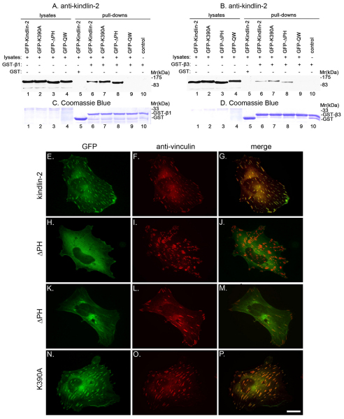
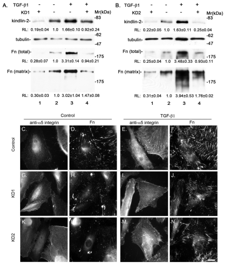
Kindlin-2 is a FERM and PH domain-containing integrin-binding protein that is emerging as an important regulator of integrin activation. How kindlin-2 functions in integrin activation, however, is not known. We report here that kindlin-2 interacts with multiple phosphoinositides, preferentially with phosphatidylinositol 3,4,5-trisphosphate. Although integrin-binding is essential for focal adhesion localization of kindlin-2, phosphoinositide-binding is not required for this process. Using biologically and clinically relevant glomerular podocytes as a model system, we show that integrin activation and dependent processes are tightly regulated by kindlin-2: depletion of kindlin-2 reduced integrin activation, matrix adhesion and fibronectin matrix deposition, whereas overexpression of kindlin-2 promoted these processes. Furthermore, we provide evidence showing that kindlin-2 is involved in phosphoinositide-3-kinase-mediated regulation of podocyte-matrix adhesion and fibronectin matrix deposition. Mechanistically, kindlin-2 promotes integrin activation and integrin-dependent processes through interacting with both integrins and phosphoinositides. TGF-β1, a mediator of progressive glomerular failure, markedly increased the level of kindlin-2 and fibronectin matrix deposition, and the latter process was reversed by depletion of kindlin-2. Our results reveal important functions of kindlin-2 in the regulation of podocyte-matrix adhesion and matrix deposition and shed new light on the mechanism whereby kindlin-2 functions in these processes.


Figures

Fig. 1.
Fig. 1.
Expression and localization of kindlin-2 in podocytes. (A) Podocytes were cultured under permissive (lane 1) or nonpermissive conditions (lane 2) for 14 days. Cell lysates were analyzed by western blotting with antibodies (Abs) recognizing kindlin-2 (2 μg proteins/lane) or p27 (10 μg proteins/lane). Equal loading was confirmed by probing the membrane with anti-tubulin Ab. (B) Lysates (45 μg proteins/lane) of podocytes (lanes 1 and 2) and HaCaT cell lysates (lane 3) were analyzed by western blotting with Abs recognizing kindlin-1 or Coomassie Blue staining. (C–H) Differentiated podocytes were dually stained with mouse anti-kindlin-2 mAb (C) and FITC-conjugated phalloidin (D) or mouse anti-kindlin-2 mAb (F) and rabbit anti-β-catenin Ab (G). The images in panels E and H were merged using the NIH ImageJ program. Scale bars: 10 μm.
Fig. 2.
Fig. 2.
Knockdown of kindlin-2 inhibits β1- and β3-integrin activation. (A,B) Differentiated podocytes were transfected with control RNA (lane 1), KD1 (A, lane 2) or KD2 (B, lane 2). The lysates (5 μg proteins/lane) were analyzed by western blotting with Abs recognizing talin, β1-integrin, β3-integrin, kindlin-2, ILK, migfilin or tubulin. Relative levels (RLs) of kindlin-2 (means ± s.d. from five independent experiments) in A and B were calculated as described in the Materials and Methods. (C) Cell adhesion to fibronectin or vitronectin was analyzed as described in the Materials and Methods. Bars represent means ± s.d. from three independent experiments. *P<0.05 versus the control. **P<0.01 versus the control. (D–G) Cell surface expression (D,E) and activation (F,G) of β1- (E,G) and β3- (D,F) integrins in kindlin-2 knockdown and control cells were analyzed as described in the Materials and Methods. Bars represent means ± s.d. from three independent experiments. *P<0.05 versus the control.
Fig. 3.
Fig. 3.
Kindlin-2 promotes integrin activation and ECM adhesion through interactions with phosphoinositides and integrins. (A) Podocytes cultured under non-permissive condition for 12 days were infected with adenoviral vectors encoding β-galactosidase (lane 1), kindlin-2 (lane 2), Q614W615AA (lane 3), ΔPH (lane 4) or K390A (lane 5) mutants. The lysates (5 μg proteins/lane) were analyzed by western blotting with Abs recognizing talin, kindlin-2, ILK, migfilin or tubulin. Relative levels (RLs) of wild-type or mutant forms of kindlin-2 (means ± s.d. from five independent experiments) were quantified as described in the Materials and Methods. (B) Cell adhesion to fibronectin was analyzed as described in the Materials and Methods. Bars represent means ± s.d. from five independent experiments. *P<0.05 versus the control. **P<0.01 versus the control. (CF) Cell surface expression (C,D) and activation (E,F) of β1- (D,F) and β3- (C,E) integrins were analyzed as described in the Materials and Methods. Bars represent means ± s.d. from four independent experiments. *P<0.05 versus the control. **P<0.01 versus the control.
Fig. 4.
Fig. 4.
Kindlin-2 interacts with phosphoinositides through the PH domain. (A) Superposition of the Akt PH domain (yellow) bound to the PI(3,4,5)P3 head group (purple; PDB1UNQ) with a model of the PH domain of kindlin-2 (light cyan) showing that the kindlin-2 PH domain K390 (red) is positioned to potentially interact with the phosphate group of the PI(3,4,5)P3 head group. Model inspection and analysis of the kindlin-2 PH domain were performed using the PYMOL program (Schwede et al., 2003). (B) GST fusion proteins containing the kindlin-2 F2 subdomain with (lane 1) or without (lane 2) the PH insert, or the GST–kindlin-2 F2 subdomain containing the PH insert bearing the K390A mutation (lane 3) were analyzed by SDS-PAGE and Coomassie Blue staining. (CE,G) PIP strips immobilized with different phospholipids (C,D,G) and a PIP array immobilized with different amounts of phosphoinositides (E) were incubated with 1 μg/ml of purified GST–F2-PH (C,E), GST–ΔPH (D) or GST–K390A (G). GST-fusion proteins bound to immobilized phosphoinositides were detected with an anti-GST Ab. (F) A representative HSQC spectrum of kindlin-2 PH domain showing D-myo-inositol 1,3,4,5-tetraphosphate and D-myo-inositol 1,4,5-triphosphate-induced chemical shift changes of the kindlin-2 PH domain. HSQC spectra were acquired as described in the Materials and Methods. Black, kindlin-2 PH domain alone; red, kindlin-2 PH domain: D-myo-inositol 1,3,4,5-tetraphosphate=1:2 (molar ratio); blue, kindlin-2 PH domain: D-myo-inositol 1,4,5-triphosphate=1:5 (molar ratio). (H) Podocyte lysates were incubated with PI(3,4,5)P3 beads (lane 3) or control beads lacking PI(3,4,5)P3 (lane 2). Kindlin-2 precipitated with PI(3,4,5)P3 beads was detected by western blotting with anti-kindlin-2 mAb. Lane 1 was loaded with 1 μg of podocyte lysates. (I) Lysates of podocytes expressing FLAG–kindlin-2, FLAG–K390A or FLAG–ΔPH were incubated with PI(3,4,5)P3 beads (lanes 5–7) or control beads lacking PI(3,4,5)P3 (lane 4) as indicated. The lysates (3 μg proteins/lane) of podocytes expressing FLAG–kindlin-2 (lane 1), FLAG–K390A (lane 2) or FLAG–ΔPH (lane 3) and proteins pulled down by PI(3,4,5)P3 beads (lanes 5–7) or the control beads (lane 4) were analyzed by western blotting with anti-kindlin-2 mAb.
Fig. 5.
Fig. 5.
Deletion of the PH domain compromises kindlin-2 clustering at FAs but phosphoinositide-binding is not essential for this process. (A–D) Human podocytes were transfected with vectors encoding GFP–kindin-2 (lanes 1, 5 and 6), GFP–K390A (lanes 2 and 7), GFP–ΔPH (lanes 3 and 8) or GFP–Q614W615AA mutant (lanes 4 and 9). A GST pull-down experiment was performed using cells expressing GFP-tagged wild-type or mutant forms of kindlin-2 as indicated in the figure. The cell lysates (2 μg proteins/lane, lanes 1–4), GST precipitates (lane 5), GST–β1 precipitates (A,C, lanes 6–9) and GST–β3 precipitates (B,D, lanes 6–9) were analyzed by western blotting with anti-kindlin-2 mAb (A,B) or Coomassie Blue staining (C,D). The samples in lane 10 were prepared as those in lanes 6–9 except cell lysates were omitted. (EP) Podocytes were transfected with vectors encoding GFP–kindlin-2 (E–G), GFP–ΔPH (H–M) or GFP–K390A (N–P). The transfectants were plated on fibronectin-coated cover slips and stained with a mouse anti-vinculin mAb and a Rhodamine-Red-conjugated anti-mouse IgG Ab. The cells were observed under a fluorescence microscope equipped with GFP (E,H,K,N) and Rhodamine (F,I,L,O) filters. The GFP and Rhodamine images were merged in panels G, J, M and P using the NIH ImageJ program. Scale bar: 10 μm.
Fig. 6.
Fig. 6.
Membrane localization of kindlin-2 and kindlin-2 PH domain. (AC) Human podocytes that were transfected with GFP–kindlin-2 vector and p110* vector (A), GFP–K390A vector and p110* vector (B) or GFP–K390A vector and a control vector lacking the PI3K sequence (C) were plated on fibronectin-coated cover slips and fixed. (D–L) Podocytes that were transfected with GFP–Akt-PH vector and p110* vector (G–L) or GFP–Akt-PH vector and a control vector lacking the PI3K sequence (D–F) were plated on fibronectin-coated cover slips, fixed and stained with anti-kindlin-2 mAb and a Rhodamine-Red-conjugated anti-mouse IgG Ab. (M–U) Podocytes that were transfected with GFP–Akt-PH vector, tdTomato-kindlin-2-PH vector and p110* vector (P–U) or GFP–Akt-PH vector, tdTomato-kindlin-2-PH vector and a control vector lacking the PI3K sequence (M–O) were plated on fibronectin-coated cover slips and fixed. Cells in J–L and S–U were treated with 0.1 μM wortmannin for 1 hour prior to fixation. Cells were observed under a fluorescence microscope equipped with GFP (A–D,G,J,M,P,S) and Rhodamine and tdTomato (E,H,K,N,Q,T) filters. The GFP and Rhodamine and tdTomato images were merged in panels F, I, L, O, R and U using the NIH ImageJ program. Arrows indicate membrane regions lacking a high concentration of GFP–Akt-PH. Arrowheads indicate membrane regions containing high concentrations of GFP–Akt-PH. Scale bar: 10 μm.
Fig. 7.
Fig. 7.
Kindlin-2 functions in TGF-β1-induced fibronectin matrix deposition in podocytes. Differentiated human podocytes were transfected with kindlin-2 siRNA KD1, kindlin-2 siRNA KD2 or control RNA and cultured in the presence or absence of TGF-β1 (5 ng/ml) as indicated in the figure. (A,B) Cell lysates and ECM fractions were analyzed by western blotting with Abs specific for kindlin-2, tubulin (as a loading control) or fibronectin as indicated in the figure. The densities of fibronectin, kindlin-2 and tubulin bands were quantified using the NIH ImageJ program. Relative levels (RLs) of kindlin-2 and fibronectin in total cell lysates or ECM fractions (means ± s.d. from three independent experiments in A and two independent experiments in B) were quantified as described in the Materials and Methods. (CN) Cells (as indicated in the figure) were dually stained with mouse anti-α5-integrin mAb SNAKA51 and rabbit anti-fibronectin Abs. The mouse and rabbit Abs were detected with secondary Rhodamine-Red-conjugated anti-mouse IgG Ab and FITC-conjugated anti-rabbit IgG Ab. The cells were observed under a fluorescence microscope equipped with Rhodamine (C,E,G,I,K,M) and FITC (D,F,H,J,L,N) filters. Scale bar: 10 μm.
Fig. 8.
Fig. 8.
The kindlin-2-phosphoinositide interaction promotes cellECM adhesion and fibronectin matrix deposition. (A) Human podocytes cultured under nonpermissive condition for 12 days were infected with adenoviral vectors encoding β-galactosidase (lane 1), kindlin-2 (lane 2), Q614W615AA (lane 3), ΔPH (lane 4) or K390A (lane 5). Kindlin-2, GAPDH (as a loading control), total cell-associated fibronectin and fibronectin in the ECM fractions were analyzed by western blotting with Abs as indicated. Relative levels (RLs) of kindlin-2 and fibronectin (means ± s.d. from two independent experiments) were calculated as described in the Materials and Methods. (B) Human podocytes were transfected with kindlin-2 siRNA KD2 (lane 1), KD1 (lane 2), control RNA (lanes 3 and 4) and a DNA vector encoding Myc-p110* (lanes 1, 2 and 4) or a control DNA vector lacking the PI3K sequence (lane 3). The cell lysates or ECM fractions were analyzed by western blotting with Abs specific for Myc, kindlin-2, Akt, phospho-Akt (Ser473), phospho-Akt (Thr308), fibronectin or GAPDH (as a loading control). Relative levels of fibronectin in total cell lysates and ECM fractions (means ± s.d. from two independent experiments) were calculated as described in the Materials and Methods. (C) Adhesion of podocytes that were transfected with control DNA vector and control RNA (control), p110* vector and control RNA (p110*), p110* vector and KD1 (p110* + KD1), and p110* vector and KD2 (p110* + KD2) to fibronectin was analyzed as described in the Materials and Methods. Error bars represent means ± s.d. from three independent experiments. *P<0.05 versus the control.

References

    1. Barisoni L., Mundel P. (2003). Podocyte biology and the emerging understanding of podocyte diseases. Am. J. Nephrol. 23, 353-360 - PubMed
    1. Byzova T. V., Goldman C. K., Pampori N., Thomas K. A., Bett A., Shattil S. J., Plow E. F. (2000). A mechanism for modulation of cellular responses to VEGF: activation of the integrins. Mol. Cell 6, 851-860 - PubMed
    1. Calderwood D. A. (2004). Integrin activation. J. Cell Sci. 117, 657-666 - PubMed
    1. Chivian D., Kim D. E., Malmstrom L., Schonbrun J., Rohl C. A., Baker D. (2005). Prediction of CASP6 structures using automated Robetta protocols. Proteins 61 Suppl. 7, 157-166 - PubMed
    1. Clark K., Pankov R., Travis M. A., Askari J. A., Mould A. P., Craig S. E., Newham P., Yamada K. M., Humphries M. J. (2005). A specific {alpha}5{beta}1-integrin conformation promotes directional integrin translocation and fibronectin matrix formation. J. Cell Sci. 118, 291-300 - PMC - PubMed

Publication types

MeSH terms