. 2011 Mar 8;108(10):3842-7.
doi: 10.1073/pnas.1013377108. Epub 2011 Feb 15.

Two ground state isoforms and a chromophore D-ring photoflip triggering extensive intramolecular changes in a canonical phytochrome

Affiliations

Two ground state isoforms and a chromophore D-ring photoflip triggering extensive intramolecular changes in a canonical phytochrome

Chen Song et al. Proc Natl Acad Sci U S A. .

Abstract

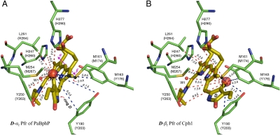
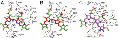
Phytochrome photoreceptors mediate light responses in plants and in many microorganisms. Here we report studies using (1)H-(13)C magic-angle spinning NMR spectroscopy of the sensor module of cyanobacterial phytochrome Cph1. Two isoforms of the red-light absorbing Pr ground state are identified. Conclusive evidence that photoisomerization occurs at the C15-methine bridge leading to a β-facial disposition of the ring D is presented. In the far-red-light absorbing Pfr state, strong hydrogen-bonding interactions of the D-ring carbonyl group to Tyr-263 and of N24 to Asp-207 hold the chromophore in a tensed conformation. Signaling is triggered when Asp-207 is released from its salt bridge to Arg-472, probably inducing conformational changes in the tongue region. A second signal route is initiated by partner swapping of the B-ring propionate between Arg-254 and Arg-222.


Conflict of interest statement

The authors declare no conflict of interest.

Figures

Fig. 1.
Fig. 1.
Interfacial 1H contacts of the PCB chromophore. (A) Schematic of protein-bound PCB shown in the Pr ZZZssa geometry. The tetrapyrrole rings and representative PCB atoms are labeled for reference. (B and C) Structural views showing residue contacts of the chromophore observed in heteronuclear correlation NMR spectra (see also Figs. S1 and S2) as (B) Pr and (C) Pfr. The presentations for Pr (B) and Pfr (C) states are modeled according to the Cph1 2VEA (21) and PaBphP 3C2W crystal structures (22), respectively. Water locations (W1–W7) are marked as red spheres and numbered (42). (DG) Close-up views of the 1H contacts of the two D-ring carbons, C19 and C171, as Pr (D and F) and Pfr (E and G). The dashed lines highlight the formula image 
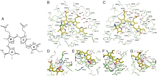
interfacial correlations during Pr → Pfr photoconversion. All 1H contacts of the chromophore are summarized in
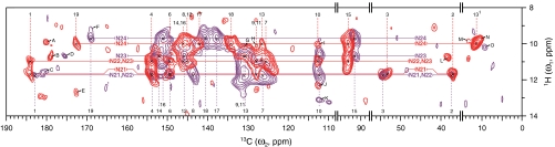
 Datasets S3 and S4.
Fig. 2.
Fig. 2.
Light-induced changes in PCB chromophore geometry. The contour plot of the 2D 1H–13C correlation spectra of u-[13C,15N]-PCB-Cph1Δ2 as Pr (red) and Pfr (purple) reveals the intramolecular proximity between protons bound to tetrapyrrole nitrogens (HN21–N24) and chromophore carbons (Dataset S1). Chemical shifts of 13C (as indicated by dashed vertical lines, Pr in red, Pfr in purple) and HN (indicated by solid horizontal lines, Pr in red, Pfr in purple) are in agreement with our previous data (18, 33). Several residue correlations in the close proximity to the chromophore (within a region of approximately 3.5 Å) are also resolved (labeled with Latin capitals A to O, listed in Dataset S2). Spectral regions without resonances, 15–35, 57–88, and 95–109 ppm in the ω2-dimension are omitted. For 1H spectra in the ω1-dimension, only the characteristic spectral region of 8–14 ppm is shown.
Fig. 3.
Fig. 3.
D-ring facial disposition in the Pfr state of PaBphP and Cph1. In the PaBphP 3C2W Pfr structure (A) (22), the light-induced photoisomerization tilts the ring D approximately 50° anticlockwise with respect to the PCB plane (rings B and C). Such an α-facial D-ring disposition (D-αf) of PaBphP (indicated by a solid arrow; ref. 30) leads to a poor agreement with our observed NMR contacts for Cph1, in particular D-ring carbons (e.g., C171, Datasets S3 and S4). Some 1H contacts of the C171 within the detection range of C–H distance (approximately 5.5 Å) are not resolved (red dashed lines, A), others appear for the C171 despite a distance much beyond the 5.5-Å prediction (blue dashed lines, A). If, on the other hand, the chromophore adopts β-facial disposition for its ring D (D-βf) relative to the rings B and C (B), the distance constraints can match our observed NMR contacts for Cph1 very well, as shown for the C171 (blue dashed lines, B). The residues codes for Cph1 sequence are given in parentheses following the equivalent codes for PaBphP sequence (see Dataset S5).
Fig. 4.
Fig. 4.
Hydrogen-bonding networks and charge distribution inside the PCB-binding pocket. (AC) Region surrounding the PCB chromophore for the two Pr isoforms (Pr-I, A and Pr-II, B) as well as for Pfr (C). Dashed lines highlight the hydrogen-bonding networks in the tetrapyrrole cavity. The + (blue) and − (red) signs represent the positive and negative charges, respectively. The conjugation pattern of the chromophore is colored in red for Pr (A and B) and purple for Pfr (C) (18).

References

    1. Butler WL, Norris KH, Siegelman HW, Hendricks SB. Detection, assay, and preliminary purification of the pigment controlling photoresponsive development of plants. Proc Natl Acad Sci USA. 1959;45:1703–1708. - PMC - PubMed
    1. Smith H. Phytochromes and light signal perception by plants—an emerging synthesis. Nature. 2000;407:585–591. - PubMed
    1. Hughes J, et al. A prokaryotic phytochrome. Nature. 1997;386:663. - PubMed
    1. Yeh K-C, Wu S-H, Murphy JT, Lagarias JC. A cyanobacterial phytochrome two-component light sensory system. Science. 1997;277:1505–1508. - PubMed
    1. Davis SJ, Vener AV, Vierstra RD. Bacteriophytochromes: Phytochrome-like photoreceptors from nonphotosynthetic eubacteria. Science. 1999;286:2517–2520. - PubMed

Publication types