Specific regulation of the chemokine response to viral hemorrhagic septicemia virus at the entry site
- PMID: 21325404
- PMCID: PMC3126238
- DOI: 10.1128/JVI.02519-10
Specific regulation of the chemokine response to viral hemorrhagic septicemia virus at the entry site
Abstract
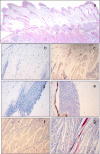
The fin bases constitute the main portal of rhabdovirus entry into rainbow trout (Oncorhynchus mykiss), and replication in this first site strongly conditions the outcome of the infection. In this context, we studied the chemokine response elicited in this area in response to viral hemorrhagic septicemia virus (VHSV), a rhabdovirus. Among all the rainbow trout chemokine genes studied, only the transcription levels of CK10 and CK12 were significantly upregulated in response to VHSV. As the virus had previously been shown to elicit a much stronger chemokine response in internal organs, we compared the effect of VHSV on the gills, another mucosal site which does not constitute the main site of viral entry or rhabdoviral replication. In this case, a significantly stronger chemokine response was triggered, with CK1, CK3, CK9, and CK11 being upregulated in response to VHSV and CK10 and CK12 being down-modulated by the virus. We then conducted further experiments to understand how these different chemokine responses of mucosal tissues could correlate with their capacity to support VHSV replication. No viral replication was detected in the gills, while at the fin bases, only the skin and the muscle were actively supporting viral replication. Within the skin, viral replication took place in the dermis, while viral replication was blocked within epidermal cells at some point before protein translation. The different susceptibilities of the different skin layers to VHSV correlated with the effect that VHSV has on their capacity to secrete chemotactic factors. Altogether, these results suggest a VHSV interference mechanism on the early chemokine response at its active replication sites within mucosal tissues, a possible key process that may facilitate viral entry.
Figures


Similar articles
-
Chemokine transcription in rainbow trout (Oncorhynchus mykiss) is differently modulated in response to viral hemorrhagic septicaemia virus (VHSV) or infectious pancreatic necrosis virus (IPNV).Fish Shellfish Immunol. 2009 Dec;27(6):661-9. doi: 10.1016/j.fsi.2009.08.003. Epub 2009 Aug 21. Fish Shellfish Immunol. 2009. PMID: 19699800
-
Transcriptomic responses in rainbow trout gills upon infection with viral hemorrhagic septicemia virus (VHSV).Dev Comp Immunol. 2014 May;44(1):12-20. doi: 10.1016/j.dci.2013.11.006. Epub 2013 Nov 22. Dev Comp Immunol. 2014. PMID: 24269609
-
Early activation of teleost B cells in response to rhabdovirus infection.J Virol. 2015 Feb;89(3):1768-80. doi: 10.1128/JVI.03080-14. Epub 2014 Nov 19. J Virol. 2015. PMID: 25410870 Free PMC article.
-
Early immune responses in rainbow trout liver upon viral hemorrhagic septicemia virus (VHSV) infection.PLoS One. 2014 Oct 22;9(10):e111084. doi: 10.1371/journal.pone.0111084. eCollection 2014. PLoS One. 2014. PMID: 25338079 Free PMC article.
-
Viral replication in excised fin tissues (VREFT) corresponds with prior exposure of Pacific herring, Clupea pallasii (Valenciennes), to viral haemorrhagic septicaemia virus (VHSV).J Fish Dis. 2011 Jan;34(1):3-12. doi: 10.1111/j.1365-2761.2010.01210.x. Epub 2010 Dec 1. J Fish Dis. 2011. PMID: 21118270
Cited by
-
A Review of the Immunological Mechanisms Following Mucosal Vaccination of Finfish.Front Immunol. 2015 Aug 24;6:427. doi: 10.3389/fimmu.2015.00427. eCollection 2015. Front Immunol. 2015. PMID: 26379665 Free PMC article. Review.
-
CK12a, a CCL19-like Chemokine That Orchestrates both Nasal and Systemic Antiviral Immune Responses in Rainbow Trout.J Immunol. 2017 Dec 1;199(11):3900-3913. doi: 10.4049/jimmunol.1700757. Epub 2017 Oct 23. J Immunol. 2017. PMID: 29061765 Free PMC article.
-
Unraveling the Systemic and Local Immune Response of Rainbow Trout (Oncorhynchus mykiss) to the Viral Hemorrhagic Septicemic Virus.Biology (Basel). 2025 Aug 5;14(8):1003. doi: 10.3390/biology14081003. Biology (Basel). 2025. PMID: 40906234 Free PMC article.
-
Immunological characterization of the teleost adipose tissue and its modulation in response to viral infection and fat-content in the diet.PLoS One. 2014 Oct 21;9(10):e110920. doi: 10.1371/journal.pone.0110920. eCollection 2014. PLoS One. 2014. PMID: 25333488 Free PMC article.
-
CD38 Defines a Subset of B Cells in Rainbow Trout Kidney With High IgM Secreting Capacities.Front Immunol. 2021 Nov 30;12:773888. doi: 10.3389/fimmu.2021.773888. eCollection 2021. Front Immunol. 2021. PMID: 34917087 Free PMC article.
References
-
- Alcami A. 2003. Viral mimicry of cytokines, chemokines and their receptors. Nat. Rev. Immunol. 3:36–50 - PubMed
-
- Alfano M., Poli G. 2005. Role of cytokines and chemokines in the regulation of innate immunity and HIV infection. Mol. Immunol. 42:161–182 - PubMed
-
- Baird J. W., et al. 1999. ESkine, a novel beta-chemokine, is differentially spliced to produce secretable and nuclear targeted isoforms. J. Biol. Chem. 274:33496–33503 - PubMed
-
- Brudeseth B. E., Castric J., Evensen O. 2002. Studies on pathogenesis following single and double infection with viral hemorrhagic septicemia virus and infectious hematopoietic necrosis virus in rainbow trout (Oncorhynchus mykiss). Vet. Pathol. 39:180–189 - PubMed
-
- Buchmann K. 1998. Histochemical characteristics of Gyrodactylus derjavini parasitizing the fins of rainbow trout (Oncorhynchus mykiss). Folia Parasitol. (Praha) 45:312–318 - PubMed
Publication types
MeSH terms
Substances
LinkOut - more resources
Full Text Sources
Other Literature Sources
Research Materials

