. 2011 Feb 16;31(7):2511-25.
doi: 10.1523/JNEUROSCI.5245-10.2011.

Tau-induced defects in synaptic plasticity, learning, and memory are reversible in transgenic mice after switching off the toxic Tau mutant

Affiliations

Tau-induced defects in synaptic plasticity, learning, and memory are reversible in transgenic mice after switching off the toxic Tau mutant

Astrid Sydow et al. J Neurosci. .

Abstract

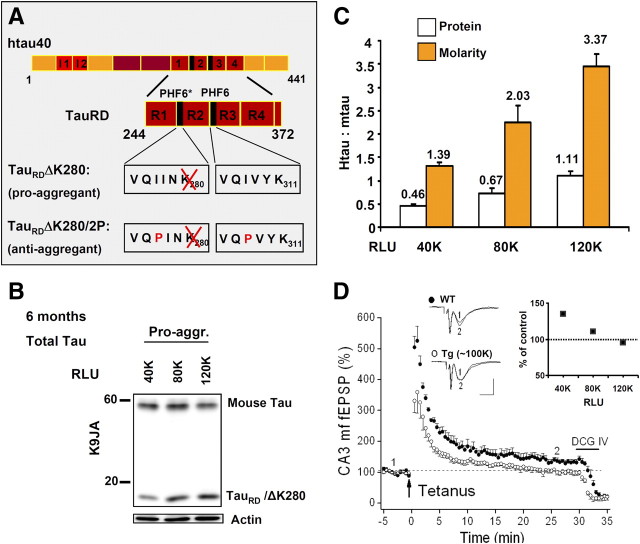
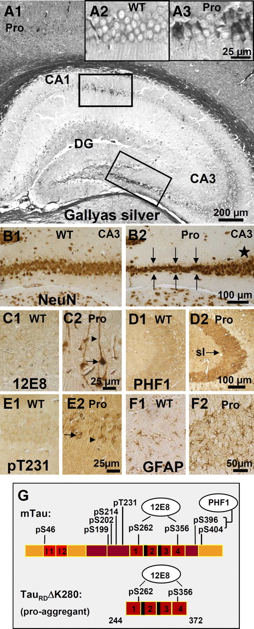
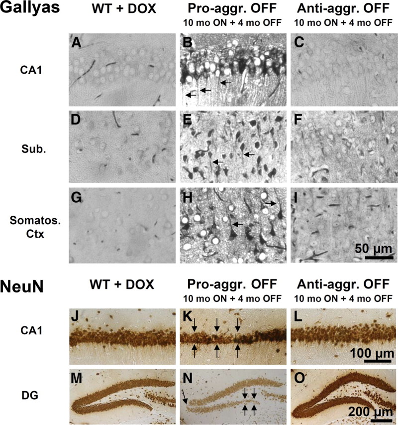
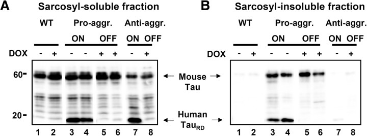
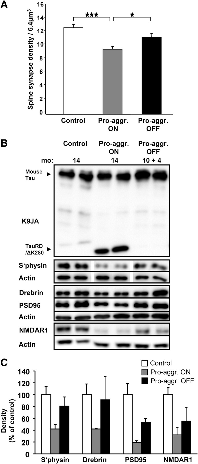
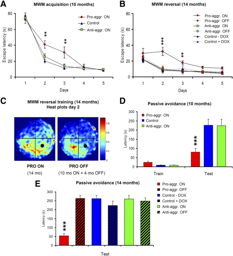
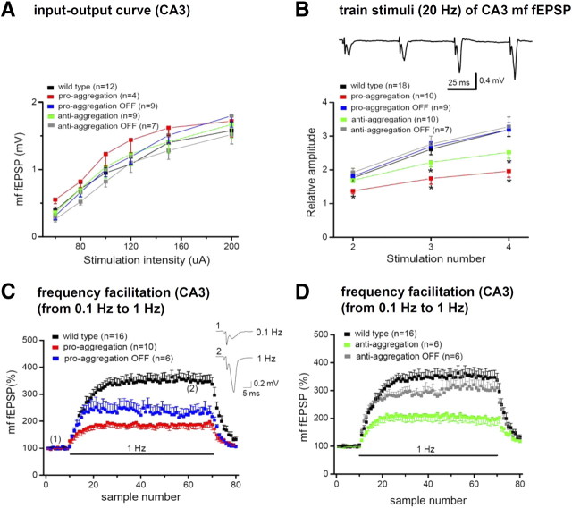
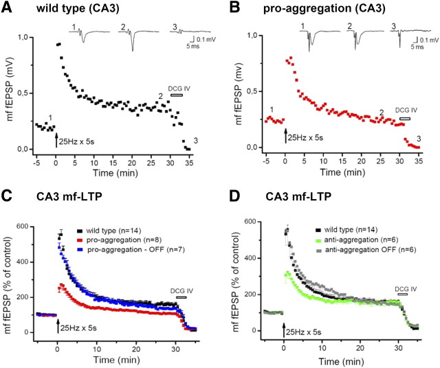
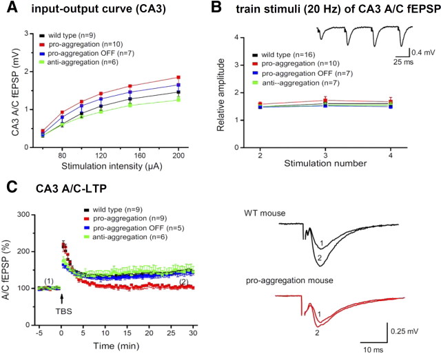
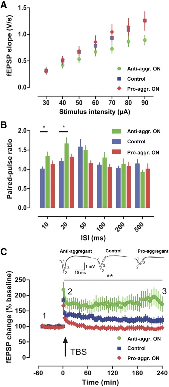
This report describes the behavioral and electrophysiological analysis of regulatable transgenic mice expressing mutant repeat domains of human Tau (Tau(RD)). Mice were generated to express Tau(RD) in two forms, differing in their propensity for β-structure and thus in their tendency for aggregation ("pro-aggregant" or "anti-aggregant") (Mocanu et al., 2008). Only pro-aggregant mice show pronounced changes typical for Tau pathology in Alzheimer's disease (aggregation, missorting, hyperphosphorylation, synaptic and neuronal loss), indicating that the β-propensity and hence the ability to aggregate is a key factor in the disease. We now tested the mice with regard to neuromotor parameters, behavior, learning and memory, and synaptic plasticity and correlated this with histological and biochemical parameters in different stages of switching Tau(RD) on or off. The mice are normal in neuromotor tests. However, pro-aggregant Tau(RD) mice are strongly impaired in memory and show pronounced loss of long-term potentiation (LTP), suggesting that Tau aggregation specifically perturbs these brain functions. Remarkably, when the expression of human pro-aggregant Tau(RD) is switched on for ∼ 10 months and off for ∼ 4 months, memory and LTP recover, whereas aggregates decrease moderately and change their composition from mixed human plus mouse Tau to mouse Tau only. Neuronal loss persists, but synapses are partially rescued. This argues that continuous presence of amyloidogenic pro-aggregant Tau(RD) constitutes the main toxic insult for memory and LTP, rather than the aggregates as such.


Figures

Figure 1.
Figure 1.
Characterization of TauRD expression in mice. A, Bar diagrams illustrating the domains of full-length Tau (2N4R, 441 residues) and the repeat domain construct K18. Black segments indicate the hexapeptide motifs responsible for aggregation via β-structure. The pro-aggregant FTDP17 mutation ΔK280 and the anti-aggregant proline mutations (I277P, I308P) are highlighted. B, Expression level of TauRD/ΔK280 versus luciferase expression (RLU). Three mice with 40,000; 80,000; and 120,000 RLU are compared. The endogenous mouse Tau (Mr ∼ 55 kDa) is roughly comparable (top), as well as the actin control (bottom). The band of TauRD (Mr ∼ 14 kDa, middle) increases with the luciferase level. Tau bands are revealed by blotting with antibody K9JA. pro-aggr., Pro-aggregant. C, Quantitation of B (n = 4). White bars indicate that the protein ratio hTau:mTau deduced from blots varies almost linearly with luciferase readings. Orange bars show corresponding molar ratios calculated on the basis of molecular masses, ranging up to ∼3.4 at an RLU value of 120,000. D, Inverse relationship between loss of CA3–mossy fiber LTP and luciferase expression (RLU). This effect occurs only with pro-aggregant TauRD. The diagram depicts normalized mossy fiber fEPSPs before and after tetanic stimulation. In wild-type (WT) hippocampi, LTP attained 130 ± 4% (filled circles; n = 4), whereas transgenic hippocampi displaying 80,000–120,000 RLU failed to exhibit significant LTP (open circles; 105 ± 6%, n = 4). Representative fEPSP recordings from wild-type and transgenic hippocampi taken at the equivalent time points of the diagram are illustrated above. The inset depicts the inverse relationship between loss of LTP and RLU (40,000, n = 3; 80,000, n = 2; 120,000, n = 2). Note that the size of LTP at 40,000 RLU (138 ± 5%, n = 3) was not significantly different from the control.
Figure 2.
Figure 2.
Histology of a pro-aggregant TauRD mouse at 10 months of expression. A, Gallyas silver staining of the hippocampus. Note prominent Tau aggregation in CA1, CA3, and DG regions of the pro-aggregant mutant mouse (A1). Insets show the boxed CA1 region with higher magnification for a Gallyas-negative wild-type (WT) animal (A2) compared with a pro-aggregant mutant mouse (A3). B, NeuN staining of neurons in the CA3 region (lower boxed area in A1) of a WT (B1) and a pro-aggregant mutant (B2) mouse. Note the loss of neurons in the pro-aggregant animal (B2, arrows) compared with the normal number of neurons in WT (B1) and the pro-aggregant mutant mouse within another part of the CA3 region (B2, star). C, Staining of somatosensoric cortical neurons by antibody 12E8 (against phosphorylated KXGS motifs inside the Tau repeat domain) for WT (C1) and pro-aggregant mutant (C2) mice. The phosphorylation (12E8 epitope) and missorting of Tau into the apical dendrites (arrowheads) and soma (arrows) appears only in the pro-aggregant mutant animal (C2). D, Pronounced PHF1 immunoreactivity (pS396, pS404) of the mossy fibers in the CA3 region of a pro-aggregant mutant mouse (D2, arrow; sl, stratum lucidum) compared with negatively stained mossy fibers of a WT mouse (D1). E, Stained CA1 neurons by an antibody against phosphorylated T231-Tau and the identification of missorted Tau into the soma (arrow) and apical dendrites (arrowhead) in the pro-aggregant mutant mouse (E2) compared with negative pyramidal neurons of the WT animal (E1). F, Staining by antibody against GFAP in the DG, revealing inflammatory processes by activated astrocytes for the pro-aggregant mutant mouse (F2) but not for the WT animal (F1). G, Bar diagram illustrating positions of phosphoepitopes inside and outside the Tau repeat domain. Staining with antibodies pS46, pS199, pS202, pS214, pT231, pS404, PHF1, and MC1 is positive, and staining with AT8 and AT270 is negative (data only partly shown) (see also Mocanu et al.,2008). Pro, Pro-aggregant mutant mouse; WT, Wild-type mouse. Scale bars: A1, 200 μm; B1, B2, D1, D2, 100 μm; F1, F2, 50 μm; A2, A3, C1, C2, E1, E2, 25 μm.
Figure 3.
Figure 3.
Histology of pro-aggregant and anti-aggregant TauRD mice at 10 months switched ON plus 4 months OFF compared with wild-type mice. Top, Gallyas silver staining for Tau aggregates. Bottom, NeuN staining for neurons. Images B, E, and H illustrate Gallyas-positive neurons in the CA1 region, subiculum, and somatosensoric cortex of pro-aggregant OFF mice compared with wild-type mice (A, D, G) and anti-aggregant OFF mice (C, F, I). Note the missorting of Tau into the apical dendrites of the neurons in pro-aggregant OFF mice (B, E, H, arrows). Images K and N show the neuronal loss in the CA1 region and DG of pro-aggregant OFF mice (arrows) compared with wild-type (J, M) and anti-aggregant OFF (L, O) mice. Note shrinkage of entire pyramidal cell layer in pro-aggregant OFF mice (K) in contrast to wild-type (J) and anti-aggregant OFF (L) mice. Pro-aggr., Pro-aggregant; Anti-aggr., anti-aggregant; WT, wild type; DOX, doxycycline; Sub., subiculum; Somatos. Ctx, somatosensoric cortex. Scale bars: A–I, 50 μm; J–L, 100 μm; M–O, 200 μm.
Figure 4.
Figure 4.
Analysis of soluble and aggregated Tau. Pro-aggregant or anti-aggregant TauRD were expressed continuously for 14 months (ON mice) or for 10 months plus 4 months off (OFF mice). The brain tissue of the mice was subjected to sarcosyl fractionation, and Tau was analyzed by blotting with antibody K9JA and compared with wild-type mice with or without doxycycline treatment. A, Sarcosyl-soluble fraction. B, Insoluble fraction. In both cases, lanes 1 and 2 show control mice without or with doxycycline treatment, lanes 3 and 4 represent mice switched on for 14 months, lanes 5 and 6 show mice switched on for 10 months and off for 4 months, and lanes 7 and 8 represent anti-aggregant mutant mice without and with doxycycline treatment. A, Lanes 3 and 4, At 14 months switch-on, the soluble fraction contains pro-aggregant TauRD, full-length mouse Tau, and some fragments. Lanes 5 and 6, After 10 months switch-on plus 4 months switch-off, the exogenous, pro-aggregant TauRD has disappeared, and only mouse Tau remains, partly fragmented. Note an approximately similar TauRD expression in anti-aggregant ON mice (lane 7) and no TauRD expression neither in wild-type (lanes 1 and 2) nor in anti-aggregant OFF (lane 8) mice. B, Lanes 3 and 4, At 14 months switch-on, the insoluble fraction of pro-aggregant ON mice contains both human TauRD and mouse Tau. In the pro-aggregant ON mice, the insoluble fraction contains ∼40% exogenous TauRD and ∼60% endogenous mouse Tau. After 10 months switch-on plus 4 months switch-off, the insoluble fraction contains no exogenous TauRD, and only mouse Tau remains (lanes 5 and 6). Note there is no detection of Tau aggregates in wild-type (lanes 1 and 2) and anti-aggregant ON and OFF (lanes 7 and 8) mice. Pro-aggr., Pro-aggregant; Anti-aggr., anti-aggregant; WT, wild type; DOX, doxycycline; −, without doxycycline; +, with doxycycline.
Figure 5.
Figure 5.
Spine-synapse density versus expression of pro-aggregant TauRD. A, Bars show quantitative evaluation of spine synapses by electron microscopy in the stratum radiatum of the CA1 hippocampal region of control mice (left), after 10 months expression of pro-aggregant TauRD (middle), and after 10 months switch-on plus 4 months switch-off (right). The synapse density decreases after 10 months of TauRD expression to 74% of control. In the case of 10 months switch-on and 4 months off, the spine synapses are rescued to 90% of control, indicating substantial recovery. bar 1/2, ★★★p = 0.000031; bar 2/3, ★p = 0.018; bar 1/3, p = 0.062. B, Western blots showing expression of Tau variants and synaptic proteins in wild-type mice (14 months old), pro-aggregant ON mice (14 months TauRD/ΔK280 expression), and pro-aggregant OFF mice (10 months ON plus 4 months OFF TauRD/ΔK280 expression). Decreased levels of PSD95, drebrin, synaptophysin, and NMDAR1 in pro-aggregant ON mice compared to wild-type mice are shown. Note that the levels of synaptic proteins are partly rescued after the switch off for 4 months. C, Densitometric analysis of the Western blots (B) for the synaptic proteins (synaptophysin, drebrin, PSD95, and NMDAR1), all normalized to β-actin (n = 4). The gray bars indicate the reduction of the synaptic proteins after TauRD/ΔK280 expression in the pro-aggregant ON mice to ∼40% compared with control mice. The black bars show the rescue of synaptic proteins in 4 months switched off pro-aggregant OFF mice to ∼70%, compared with control mice. Significance tests (two-sided t tests) resulted in p values of 0.0176 (synaptophysin), 0.0828 (drebrin), 0.0217 (PSD95), and 0.0133 (NMDAR1) for the differences between wild-type and Pro_ON, resulting in an overall p value of 0.000273 (highly significant) according to Fisher's combined probability test. Similarly, the combined p value for Pro_ON versus Pro_OFF was 0.0319 (significant), whereas the difference between wild-type and Pro-OFF was not significant (combined p value 0.203). Pro-aggr., pro-aggregant; S'physin, synaptophysin. The data in A and C are means ± SEM.
Figure 6.
Figure 6.
Pro-aggregant TauRD mice show impaired learning in the Morris water maze (MWM) and in the passive avoidance task. MWM acquisition (A, B) and probe trial (C) are shown. A, Escape latencies indicate a slower acquisition in pro-aggregant TauRD mice (red circles) compared with control (blue triangles) and anti-aggregant (green squares) mice. B, Four months later, mice were subjected to a reversal phase in which the platform was placed in another position (right). Pro-aggregant TauRD mice that had been switched on for 10 months and then switched off for 4 months (dashed line) acquired the new position at the control level, whereas pro-aggregant TauRD mice where expression of the transgene was continued were much worse at this task (full line). C, Heat plots for the pro-aggregant TauRD mice on day 2 of the reversal acquisition are shown (dwell frequency is indicated by coloration from red through blue). Pro-aggregant TauRD mice expressing TauRD (left plot) spent more time searching in the former platform quadrant (filled circles) than in the quadrant of the new platform (open circles). After suppression of pro-aggregant TauRD (10 months ON + 4 months OFF; right plot), the mice had a clear preference for the new target quadrant. D, Mean latencies during training and test trial in the passive avoidance task. Switched-ON pro-aggregant TauRD mice were impaired in retention of the single trial task as they reentered the dark compartment much faster during the retention trial compared with anti-aggregant and control mice. E, Mean latencies during the test trial in the passive avoidance task of a second cohort of mice, where the animals were separated into six groups from the beginning of the experiment. Half of the mice received doxycycline for a period of 4 months (pro-aggregant OFF, anti-aggregant OFF, and control plus Dox), whereas the rest of the mice were allowed to continue their TauRD expression. The passive avoidance task demonstrates an impaired retention for the pro-aggregant ON mice, whereas the pro-aggregant OFF mice behave as well as wild-type and anti-aggregant ON and OFF mice. All data are means ± SEM. Asterisks in A and D indicate the significance of differences between PRO-ON versus ANTI-ON and control mice (**p < 0.01; ***p < 0.001). Asterisks in B and E indicate the significance of differences between PRO-ON versus PRO-OFF, ANTI-ON, ANTI-OFF, and control mice (**p < 0.01; ***p < 0.001). Pro-aggr., Pro-aggregant; Anti-aggr., anti-aggregant.
Figure 7.
Figure 7.
Reduced short-term plasticity at the mossy fiber–CA3 synapse in pro-aggregant TauRD mice. A, Amplitudes of mossy fiber (mf) fEPSPs are plotted as a function of stimulus intensity to determine input–output relationships. B, The top trace depicts mossy fiber fEPSPs in response to four stimuli at 20 Hz recorded from a control slice. The diagram below displays the normalized fEPSP amplitude relative to the amplitude of the first fEPSP. *p < 0.05. C, Strongly reduced frequency facilitation induced by continuous stimulation at 1 Hz for 1 min in pro-aggregant mice. fEPSP amplitude were normalized to baseline value obtained at 0.1 Hz. Only partial recovery was observed in the pro-aggregant-OFF group. Inset, Individual fEPSPs from a control slice before and during frequency facilitation recorded at the same time points as in the normalized diagram. D, Reduction of frequency facilitation in anti-aggregant mice was reversed in the anti-aggregant-OFF group.
Figure 8.
Figure 8.
Cessation of pro-aggregant TauRD expression reverses the decrease of PTP and LTP at the mossy fiber–CA3 synapse. A, B, Characteristic time course of fEPSP changes after an LTP-inducing stimulation protocol in a control slice and in a slice from pro-aggregant TauRD mice. Traces above the diagram depict individual fEPSPs taken before induction of LTP, 25 min after induction and in the presence of DCG IV. C, The summary diagram demonstrates the complete reversal of LTP suppression in the pro-aggregant-OFF group. D, Impaired PTP in slices of the anti-aggregant group is reversed after switch-OFF. mf, Mossy fiber.
Figure 9.
Figure 9.
Switching off pro-aggregant TauRD expression reverses impairment of LTP at the A/C–CA3 synapse. A, Comparison of input–output relationships between wild-type and transgenic groups did not reveal significant differences. B, Facilitation of fEPSPs by a train of stimuli. The trace above the diagram was recorded in a control slice. The diagram below displays the normalized fEPSP amplitude relative to the amplitude of the first fEPSP. C, Left, Time course of the normalized fEPSP amplitude before and after LTP-inducing stimulation. Note the lack of LTP in the pro-aggregant group and full recovery in the pro-aggregant-OFF group. Right, Superimposed fEPSPs recorded from a control slice (black traces) and from a pro-aggregant slice (red traces) taken at the same time points as in the diagram on the left. WT, Wild type.
Figure 10.
Figure 10.
Absence of hippocampal LTP in the CA1 in pro-aggregant TauRD mice. A, Field recordings in the CA1 region of the hippocampus did not reveal any differences in input/output properties (p > 0.05 for all comparisons). B, PPF, a form of short-term plasticity, was altered at the 10 and 20 ms interval in anti-aggregant mice (green bars) compared with control (blue bars; *p < 0.05). C, LTP induced by a single tetanus revealed differences between the genotypes (**p < 0.01). Whereas slices of control mice developed an LTP that slowly returned to baseline during the 4 h recording period, pro-aggregant TauRD mice were completely devoid of LTP. The only potentiation obtained was PTP 1 min after tetanization. Anti-aggregant TauRD mice developed a robust LTP that was even more pronounced than that of controls. Anti-aggr., Anti-aggregant; Pro-aggr., pro-aggregant; ISI, interstimulus interval; TBS, theta burst stimulation.

Similar articles

Cited by

References

    1. Allen B, Ingram E, Takao M, Smith MJ, Jakes R, Virdee K, Yoshida H, Holzer M, Craxton M, Emson PC, Atzori C, Migheli A, Crowther RA, Ghetti B, Spillantini MG, Goedert M. Abundant tau filaments and nonapoptotic neurodegeneration in transgenic mice expressing human P301S tau protein. J Neurosci. 2002;22:9340–9351. - PMC - PubMed
    1. Arendash GW, Lewis J, Leighty R, McGowan E, Cracchiolo JR, Hutton M, Garcia MF. Multi-metric behavioral comparison of APPsw and P301L models for Alzheimer's disease: linkage of poorer cognitive performance to tau pathology in forebrain. Brain Res. 2004;1012:29–34. - PubMed
    1. Ballatore C, Lee VM, Trojanowski JQ. Tau-mediated neurodegeneration in Alzheimer's disease and related disorders. Nat Rev Neurosci. 2007;8:663–672. - PubMed
    1. Balschun D, Wolfer DP, Bertocchini F, Barone V, Conti A, Zuschratter W, Missiaen L, Lipp HP, Frey JU, Sorrentino V. Deletion of the ryanodine receptor type 3 (RyR3) impairs forms of synaptic plasticity and spatial learning. EMBO J. 1999;18:5264–5273. - PMC - PubMed
    1. Barghorn S, Zheng-Fischhöfer Q, Ackmann M, Biernat J, von Bergen M, Mandelkow EM, Mandelkow E. Structure, microtubule interactions, and paired helical filament aggregation by tau mutants of frontotemporal dementias. Biochemistry. 2000;39:11714–11721. - PubMed

Publication types

MeSH terms