. 2011 Mar 15;186(6):3606-14.
doi: 10.4049/jimmunol.1003470. Epub 2011 Feb 16.

A meningococcal factor H binding protein mutant that eliminates factor H binding enhances protective antibody responses to vaccination

Affiliations

A meningococcal factor H binding protein mutant that eliminates factor H binding enhances protective antibody responses to vaccination

Peter T Beernink et al. J Immunol. .

Abstract

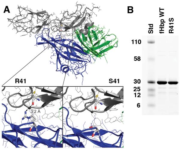
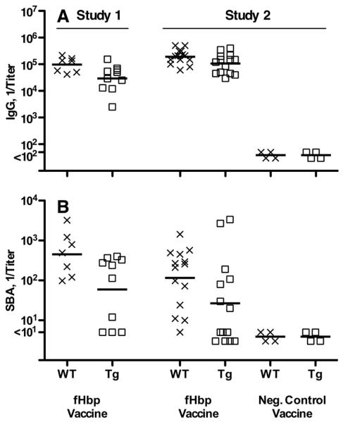
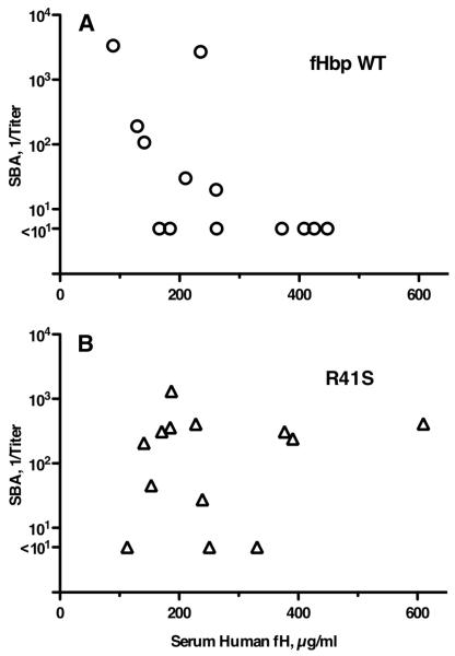
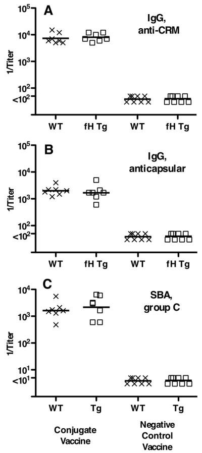
Certain pathogens recruit host complement inhibitors such as factor H (fH) to evade the immune system. Microbial complement inhibitor-binding molecules can be promising vaccine targets by eliciting Abs that neutralize this microbial defense mechanism. One such Ag, meningococcal factor H-binding protein (fHbp), was used in clinical trials before the protein was discovered to bind fH. The potential effect of fH binding on vaccine immunogenicity had not been assessed in experimental animals because fHbp binds human fH specifically. In this study, we developed a human fH transgenic mouse model. Transgenic mice immunized with fHbp vaccine had 4- to 8-fold lower serum bactericidal Ab responses than those of control mice whose native fH did not bind the vaccine. In contrast, Ab responses were unimpaired in transgenic mice immunized with a control meningococcal group C polysaccharide-protein conjugate vaccine. In transgenic mice, immunization with an fH nonbinding mutant of fHbp elicited Abs with higher bactericidal activity than that of fHbp vaccination itself. Abs elicited by the mutant fHbp more effectively blocked fH binding to wild-type fHbp than Abs elicited by fHbp that bound fH. Thus, a mutant fHbp vaccine that does not bind fH but that retains immunogenicity is predicted to be superior in humans to an fHbp vaccine that binds human fH. In the case of mutant fHbp vaccination, the resultant Ab responses may be directed more at epitopes in or near the fH binding site, which result in greater complement-mediated serum bactericidal activity; these epitopes may be obscured when human fH is bound to the wild-type fHbp vaccine.


Figures

Figure 1
Figure 1
A, Structural model of fHbp bound to a fragment of fH based on published atomic coordinates (43). The blue and green ribbons represent the respective N- and C-terminal domains of the fHbp molecule. The gray ribbon represents the sixth and seventh short consensus repeat domains of human fH previously shown to mediate the interaction of human fH and fHbp (43). The arginine residue at position 41 formed a charged H-bond with fH (left lower inset), which was predicted to be eliminated when arginine was replaced by serine (right lower inset). B, SDS-PAGE of fHbp. Wild-type (WT) fHbp and R41S mutant fHbp (5 μg/lane) were separated on 4-12% polyacyrlamide gradient gel (NuPAGE, Invitrogen, Carlsbad, CA) and stained with Coomassie blue. The molecular weights (in kDa) of the protein standards (Std) are labeled on the left.
Figure 2
Figure 2
Characterization of fHbp vaccine antigens. A, By ELISA, the fHbp WT (blue circles) bound fH, whereas the R41S substitution (red triangles, dashed line) eliminated binding of soluble human fH to solid-phase mutant fHbp. B, Binding of anti-fHbp MAb JAR 4 to solid-phase wild-type or R41S mutant fHbp indicated that similar amounts of the two proteins were adsorbed to the wells of the microtiter plate and that a conformational epitope in the N-terminal domain was retained. Symbols are the same as used in panel A. The data in panels A and B represent the mean and SE of three to six independent measurements. For data points without apparent error bars, the variability was too small to be evident. C, Binding of soluble fHbp to immobilized human fH as measured by surface plasmon resonance. Human fH (4000 response units) was coupled to the biosensor chip; the WT fHbp (0.5 μM, solid black line) showed +77.4 response units while 0.5 (solid orange line) or 1.0 μM (solid purple line) of the mutant R41S fHbp showed no binding (−1.0 and −0.8, respectively). Non-specific binding of the mutant fHbp is evident at 2.5 (red dotted line) or 5.0 (green dashed line) μM. D, Thermal stability of WT (solid black line) and R41S (dashed red line) proteins measured by differential scanning calorimetry. Protein solutions (0.5 mg/ml) were in PBS and the scan rate was 60 °C/h. Reference buffer data were subtracted as a baseline and the data were normalized based on the calculated molecular weight of the recombinant fHbp (27.7 kDa). The lower and higher temperature transitions correspond to the unfolding of the N- and C-terminal domains, respectively (46).
Figure 3
Figure 3
Serum bactericidal responses of wild-type mice to fHbp vaccines. Titers of individual mice immunized with different vaccines are represented by symbols (fHbp WT vaccine, circles; R41S mutant vaccine, triangles; and E218A/E239A mutant vaccine, + symbols). The geometric mean titers are indicated by horizontal lines. In an initial study (left), wild-type mice were immunized with three doses of fHbp WT or R41S mutant vaccines. As a control, a group of mice were immunized with a second mutant fHbp vaccine, E218A/E239A, which in previous studies showed lack of fH binding but had impaired immunogenicity in wild-type mice (30). Mice immunized with the R41S mutant developed similar geometric mean bactericidal titers as those immunized with the wild-type fHbp (71 vs. 77, respectively, P=0.32), whereas mice immunized with the E218A/E239A mutant had a GMT of 9 (P=0.01, compared to wild-type fHbp vaccine). The similar immunogenicity of the wild-type fHbp and R41S vaccines was confirmed in a replicate study, which is shown on the right (GMTs of 115 vs. 83, respectively, P=0.90).
Figure 4
Figure 4
Generation of human factor H transgenic mice. A. Identification of human factor H transgenic mice by PCR analysis of genomic DNA from tails. Amplified product (232 bp) was from human factor H transgenic mice (lane 1), wild-type mice (lane 2), and plasmid containing human factor H cDNA (lane 3). B. Human factor H detected in sera from transgenic mice. Human fH visualized by Western blotting. Serum from BALB/c mice that expressed human fH (Lane 1), wild-type BALB/c mice negative for human fH (Lane 2), and normal human serum (Lane 3) were separated on a 4-12% polyacrylamide gel by electrophoresis; proteins were transferred to a PVDF membrane. Membranes were probed with affinity isolated goat anti-human fH (Complement Technology Inc., Tyler, TX) that recognized human, but not mouse, fH (48). C. Human serum fH quantification by ELISA. Each individual serum sample is represented by a single data point of the mean concentration from two to three independent measurements. Positive mice (square symbols, N=39) had human fH concentrations >90 μg/ml and negative mice (X symbols, N=26) had concentrations <12 μg/ml, which was the lower limit of detection in the assay. For comparison, concentrations of human fH were measured in stored sera from 25 healthy adult humans (diamond symbols).
Figure 5
Figure 5
Serum antibody responses of wild-type (WT, X symbols) or human fH transgenic (Tg, square symbols) mice immunized with a fHbp vaccine that bound human fH. A, IgG anti-fHbp responses. Titers of individual mice are represented by the respective symbols and the geometric mean titers are indicated by horizontal lines. The symbols for mice immunized with negative control vaccines are titers of serum pools, each from 3-4 mice. B, Serum bactericidal responses measured with human complement against group B strain H44/76. Symbols represent titers of individual mice as in Panel A. Comparing respective IgG GMT of fHbp immunized WT and Tg mice, Study 1, P=0.03; Study 2, P=0.025. Comparing respective SBA GMT of fHbp immunized WT and Tg mice, Study 1, P=0.03; Study 2, P=0.05.
Figure 6
Figure 6
Relationship of serum group B bactericidal antibody (SBA) titers and serum human fH concentrations of transgenic mice. A, Tg mice immunized with the fHbp wild-type (WT, circular symbols) vaccine that bound fH (r= −0.65, P = 0.02, Pearson correlation coefficient between log10 SBA and log10 fH). B, Tg mice immunized with the fHbp R41S mutant vaccine (triangular symbols) that did not bind fH (r= +0.17, P= 0.58). The respective r values were significantly different (P=0.03, performed as described (42)).
Figure 7
Figure 7
Serum antibody responses of wild-type (WT, X symbols) and human fH transgenic (fH Tg, square symbols) mice immunized in Study 2 with a control meningococcal group C polysaccharide-diphtheria CRM197 conjugate vaccine. A, IgG anti-diphtheria CRM197 antibody titers (IgG anti-CRM). B, IgG group C anticapsular antibody titers. C, serum bactericidal titers (SBA) against group C strain 4243. Each symbol represents the titer of an individual mouse. Horizontal lines represent reciprocal geometric mean titers. Negative controls included serum pools from wild-type (N=8) or transgenic mice (N=8) immunized with adjuvant only or an irrelevant antigen vaccine.
Figure 8
Figure 8
Serum antibody responses of human fH transgenic mice immunized in Study 2 with a mutant fHbp (R41S) vaccine that did not bind fH. A, IgG anti-fHbp responses. Titers of individual mice are represented by symbols (control fHbp WT vaccine, circles; R41S mutant vaccine, triangles; Al(OH)3 control, filled diamonds). The geometric mean titers are indicated by horizontal lines. The symbols for mice immunized with negative control vaccine are from testing serum pools (each pool from 3-4 individual mice). Comparing respective IgG GMT of wild-type fHbp and R41S mutant vaccine groups, P=0.96. B, Serum bactericidal responses measured with human complement against group B strain H44/76. Comparing respective SBA GMT of wild-type fHbp and R41S mutant vaccine groups, P=0.11. When the analysis of the SBA responses was adjusted by general linear regression for the confounding inverse effect of serum human fH concentrations on SBA responses of Tg mice immunized with the fHbp that bound fH, the higher responses to the mutant R41S vaccine were significant (P=0.018, See Figure 9 and text).
Figure 9
Figure 9
Effect of serum human fH concentrations on ratio of bactericidal antibody responses elicited by R41S mutant fHbp vaccine versus wild-type fHbp vaccine. The respective GMT ratios (R41S mutant : fHbp wild-type) were estimated from the general linear regression model (see text), which showed that the effect of fHbp vaccine type differed by serum fH concentration on bactericidal titer (P=0.018). Based on the regression model, the ratios of the geometric mean bactericidal responses of the group immunized with R41S fHbp vaccine over that of the group immunized with wild-type fHbp vaccine were significantly greater than 1 (in favor of the mutant fHbp vaccine) for all human fH concentrations >250 μg/ml (P<0.05), and for >316 μg/ml (P<0.01).
Figure 10
Figure 10
A, Inhibition of binding human fH to fHbp by immune sera as measured by ELISA. Dilutions of individual sera from transgenic mice (N=11 per vaccine group) immunized with wild-type (circular symbols) or R41S mutant (triangular symbols) fHbp vaccines were tested for their ability to inhibit binding of fH present in 5% IgG-depleted human serum (used as a source of human fH) to immobilized wild-type fHbp. At 1:100 and 1:400 dilutions, there was significantly greater inhibitory activity in the R41S mutant fHbp vaccine group (open triangles) than mice given the fHbp vaccine that bound fH (open circles, P<0.03). Filled diamonds represent inhibition of serum pools from transgenic mice immunized with aluminum adjuvant alone (N=4 pools). B, Correlation between fH inhibition and serum bactericidal activity of individual sera. Data are from sera of individual transgenic mice immunized with wild-type or R41S mutant fHbp vaccines (r = +0.69, P=0.0004).

References

    1. Serruto D, Rappuoli R, Scarselli M, Gros P, van Strijp JA. Molecular mechanisms of complement evasion: learning from staphylococci and meningococci. Nat Rev Microbiol. 2010;8:393–399. - PubMed
    1. Meri S, Jordens M, Jarva H. Microbial complement inhibitors as vaccines. Vaccine. 2008;26(Suppl 8):I113–117. - PubMed
    1. Fearon DT, Austen KF. Activation of the alternative complement pathway due to resistance of zymosan-bound amplification convertase to endogenous regulatory mechanisms. Proc Natl Acad Sci U S A. 1977;74:1683–1687. - PMC - PubMed
    1. Weiler JM, Daha MR, Austen KF, Fearon DT. Control of the amplification convertase of complement by the plasma protein beta1H. Proc Natl Acad Sci U S A. 1976;73:3268–3272. - PMC - PubMed
    1. Pangburn MK, Schreiber RD, Muller-Eberhard HJ. Human complement C3b inactivator: isolation, characterization, and demonstration of an absolute requirement for the serum protein beta1H for cleavage of C3b and C4b in solution. J Exp Med. 1977;146:257–270. - PMC - PubMed

Publication types

MeSH terms