. 2011 Apr 15;22(8):1191-206.
doi: 10.1091/mbc.E10-07-0599. Epub 2011 Feb 16.

Mitotic progression becomes irreversible in prometaphase and collapses when Wee1 and Cdc25 are inhibited

Affiliations

Mitotic progression becomes irreversible in prometaphase and collapses when Wee1 and Cdc25 are inhibited

Tamara A Potapova et al. Mol Biol Cell. .

Abstract

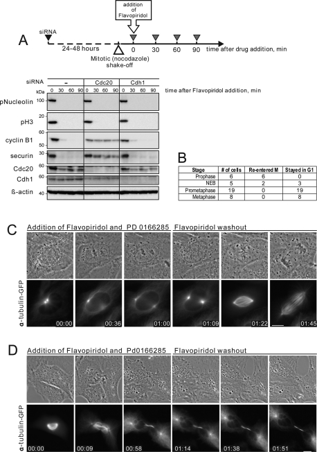
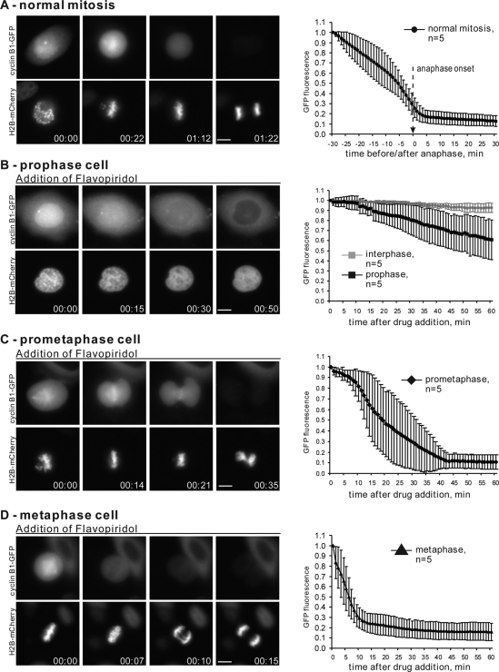
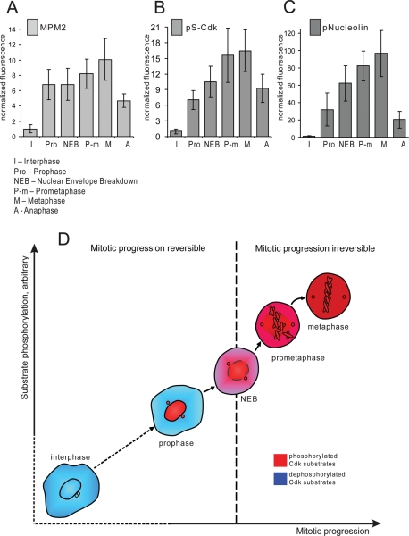
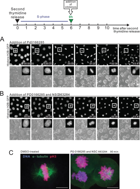
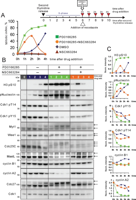
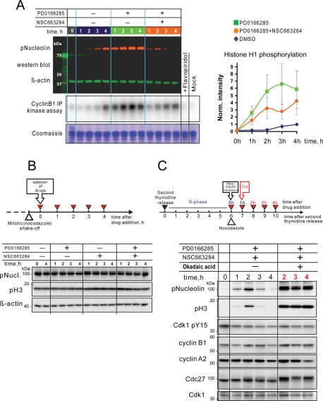
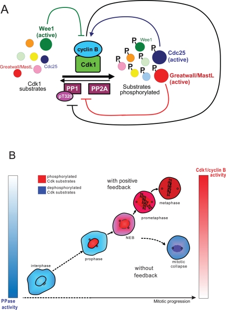
Mitosis requires precise coordination of multiple global reorganizations of the nucleus and cytoplasm. Cyclin-dependent kinase 1 (Cdk1) is the primary upstream kinase that directs mitotic progression by phosphorylation of a large number of substrate proteins. Cdk1 activation reaches the peak level due to positive feedback mechanisms. By inhibiting Cdk chemically, we showed that, in prometaphase, when Cdk1 substrates approach the peak of their phosphorylation, cells become capable of proper M-to-G1 transition. We interfered with the molecular components of the Cdk1-activating feedback system through use of chemical inhibitors of Wee1 and Myt1 kinases and Cdc25 phosphatases. Inhibition of Wee1 and Myt1 at the end of the S phase led to rapid Cdk1 activation and morphologically normal mitotic entry, even in the absence of G2. Dampening Cdc25 phosphatases simultaneously with Wee1 and Myt1 inhibition prevented Cdk1/cyclin B kinase activation and full substrate phosphorylation and induced a mitotic "collapse," a terminal state characterized by the dephosphorylation of mitotic substrates without cyclin B proteolysis. This was blocked by the PP1/PP2A phosphatase inhibitor, okadaic acid. These findings suggest that the positive feedback in Cdk activation serves to overcome the activity of Cdk-opposing phosphatases and thus sustains forward progression in mitosis.


Figures

FIGURE 1:
FIGURE 1:
Cells commit to forward mitotic progression in prometaphase. (A) Chemical Cdk inhibition in mitosis induces cyclin B1 and securin degradation that requires APC/C-Cdc20. HeLa cells were transfected with 50 nM Cdc20 siRNA for 24 h or with 50 nM Cdh1 siRNA for 48 h. Mitotic cells were collected in nocodazole, treated with 10 μM Flavopiridol for 30, 60, and 90 min, then lysed and processed for Western blotting. Depletion of Cdc20, but not Cdh1, inhibits degradation of cyclin B and securin. (B) Summary of the live imaging data from Xenopus S3 cells treated with Cdk inhibitor, Flavopiridol, and Wee1/Myt1 inhibitor, PD 0166285 at different stages of the mitotic progression. Flavopiridol was washed out 1 h after addition. (C) A prophase Xenopus S3 cell expressing alpha tubulin-GFP was treated with Cdk inhibitor, Flavopiridol, and Wee1/Myt1 inhibitor, PD 0166285. After treatment with Cdk inhibitor, mitotic progression stopped, the chromosomes decondensed, and the cell returned to an interphase morphology. Flavopiridol was washed out at 1 h, and the cell re-entered mitosis, indicating that Cdk1-activating cyclins were preserved. The cell then progressed normally trough mitosis. (D) An early prometaphase Xenopus S3 cell expressing alpha tubulin-GFP was treated with Flavopiridol and PD 0166285. After treatment at time 0, the cell underwent cytokinesis without chromosome segregation, the chromosomes decondensed, the nuclear envelope reformed, and an interphase array of microtubules appeared. Flavopiridol was washed out at 1 h. However, the cell did not re-enter mitosis, indicating that it had advanced to a G1-like state. The complete time-lapse sequences for (A) and (B) are shown in Supplemental Videos 1 and 2. Bar, 10 μm.
FIGURE 2:
FIGURE 2:
Cdk1 inhibition after prophase induces cyclin B degradation. (A) HeLa cells expressing wild-type cyclin B1-GFP and histone H2B-mCherry through normal mitosis. At left is an example of live cell imaging of a HeLa cell transiting mitosis. At right is plotted normalized cyclin B1-GFP intensity starting 30 min before anaphase onset for 5 cells. (B) Prophase, (C) prometaphase, and (D) metaphase HeLa cells expressing cyclin B1-GFP were treated with Flavopiridol at time 0. Flavopiridol induced chromosome decondensation and degradation of cyclin B. At left are examples of time-lapse imaging. On the right, normalized fluorescence intensity of the GFP is plotted starting from the time of Flavopiridol addition for five cells from each stage of mitosis. The rate of Flavopiridol-induced cyclin B degradation increases with the stage of mitosis. Complete time-lapse sequences for (A–D) are shown in Supplemental Videos 3–6. Bar, 10 μm.
FIGURE 3:
FIGURE 3:
Cdk1 phosphoepitopes rise rapidly during early mitosis. HeLa cells synchronized by double thymidine block were fixed 9 h after the release from the second thymidine block and immunolabeled with following antibodies: MPM2 (A), pS-Cdk (B), and pNucleolin (C). The fluorescence intensities were plotted according to mitotic stage. To assess the stage of mitosis precisely, cells were costained with antibody against Lamin B and with DNA dye. Integrated fluorescence intensity of a cell was measured at the brightest plane of a z series taken at 1-μm intervals. Each bar on the graphs represents an average of 15–30 cells for each stage. Error bars denote standard deviation. Representative images of each stage are shown in Supplemental Figure 2. (D) Diagram depicting relationship between Cdk substrate phosphorylation and irreversible mitotic entry. Cells become committed to forward mitotic progression in prometaphase, when Cdk substrates become phosphorylated.
FIGURE 4:
FIGURE 4:
Mitotic progression in cells synchronized at S/G2 and treated with Wee1/Myt1 and Cdc25 inhibitors. (A, B) HeLa cells stably expressing fluorescent histone H2B fused to GFP were synchronized by the double thymidine block at the S/G2 border and treated with the Wee1/Myt1 inhibitor, PD0166285, alone (A) or in combination with Cdc25 inhibitor, NSC663284 (B). While the Wee1/Myt1 inhibitor alone rapidly triggers mitosis in the majority of cells, the combination of the Wee1/Myt1 and Cdc25 inhibitors results in slow mitotic entry followed by mitotic collapse. The complete time-lapse sequence is shown in Supplemental Videos 7 and 8. Bar, 10 μm. (C) Synchronized HeLa cells were treated with the Wee1/Myt1 inhibitor, PD0166285, alone or in combination with Cdc25 inhibitor, NSC663284, for 90 min. Cells were then fixed and processed by immunofluorescence for alpha-tubulin and phosphorylated-histone H3 on S10 (mitotic marker). Labeling shows disorganized mitotic spindle, and in some cells, reduced mitotic marker.
FIGURE 5:
FIGURE 5:
Inhibition of Wee1/Myt1 and Cdc25 in synchronized cells causes mitotic collapse. (A) HeLa cells were synchronized at the S/G2 border after double thymidine block and then treated with the Wee1/Myt1 inhibitor, PD0166285, Cdc25 inhibitor, NSC663284, and the combination of the two drugs. Nocodazole was added to the medium to prevent mitotic exit. Cells were then collected at indicated time points, fixed and stained with antibody to phospho-histone H3 (mitotic marker) conjugated with Alexa Fluor 647, and processed by flow cytometry. In cells treated with vehicle only (DMSO, blue line), the mitotic index progressively increased, with more than half the cells being in mitosis by the end of the experiment. Cdc25 inhibitor, NSC663284, blocked mitotic entry (brown line). Wee1 inhibitor, PD0166285 (green line), caused rapid mitotic entry during the first hour after its addition. In cells treated with both PD0166285 and NSC663284 (orange line), the mitotic index first increased then fell. (B) HeLa cells were treated as in (A), lysed and analyzed by SDS–PAGE. In cells not treated with inhibitors (blue lanes), phosphorylations on histone H3 and nucleolin appeared by 8 h after second thymidine release and increased for the duration of the experiment. Phosphorylation of Cdk1 on inhibitory T14 and Y15 decreased over time, indicating the activation of the Cdk1/cyclin B complex. As cells were entering mitosis, a portion of Wee1, Myt1 Cdc25C, Cdc27, and MastL acquired an electrophoretic mobility shift. Cyclin B1 levels were increasing, and cyclin A2 levels dropped slightly as cells accumulated in mitosis. Inhibition of Wee1 and Myt1 kinases with PD0166285 (green lanes) resulted in rapid phosphorylation of Nucleolin and histone H3 that peaked 2 h after the drug addition and remained steadily high for the duration of the experiment. Cdk1 was rapidly dephosphorylated on inhibitory T14 and Y15. Wee1, Myt1, Cdc25, and Cdc27 rapidly shifted up. By 1 h after drug addition, Cyclin A2 was largely degraded and cyclin B1 was stable. Inhibition of Wee1 and Myt1 together with Cdc25 by addition of both PD0166285 and NSC 663284 (orange lanes) triggered the a weak phosphorylation on Nucleolin and histone H3 that peaked at 1–2 h and disappeared at 3–4 h after addition of the two drugs. Reduced mitotic phosphorylation shifts of Wee1, Myt1, Cdc25, and Cdc27 indicated that these proteins were not fully phosphorylated. Note that cyclin B and most of the cyclin A were not degraded in these cells. Panels on the right show quantifications of indicated Western blots. All values were adjusted for loading and normalized to the 4-h time point of DMSO-treated cells.
FIGURE 6:
FIGURE 6:
Deposphorylation of mitotic substrates in “collapsed” cells is a result of incomplete inhibition of Cdk-opposing phosphatases. (A) Cdk1/cyclin B1 activity does not drop in mitotic collapse cells. HeLa cells were synchronized at the S/G2 border and treated with the Wee1/Myt1 inhibitor, PD0166285, Cdc25 inhibitor, NSC663284, and the combination of the two in the presence of nocodazole. Cells were then collected at indicated time points and lysed. An aliquot of the lysate was analyzed by Western blotting for Nucleolin phosphorylation. β-Actin served as a loading control. Cyclin B1/Cdk1 complex was immunoprecipitated from the rest of the lysate and subjected to an in vitro kinase assay using histone H1 as a substrate. The kinase reaction mixture was resolved by SDS–PAGE, and the gel was exposed to phosphor-screen, which was then scanned with phosphor-imager. For a control, samples derived from the 4-h time point of DMSO-treated cells were treated with Cdk inhibitor (lane labeled “+Flavopiridol”), or processed omitting cyclin B1 antibody from immunoprecipitation (lane labeled “mock”). The gel was subsequently stained with Coomassie blue for loading. Panel on the right shows quantifications of histone H1 phosphorylation normalized to the 4 h time point of DMSO-treated cells. An average of three independent assays is shown. Error bars denote SD. (B) Simultaneous inhibition of Wee1/Myt1 and Cdc25 in cells already in mitosis does not cause mitotic substrate dephosphorylation. Mitotic HeLa cells were collected in nocodazole and then treated with Wee1/Myt1 and Cdc25 inhibitors for the indicated time, lysed, and analyzed by Western blotting. Mitotic substrates nucleolin and histone H3 remained phosphorylated throughout the experiment. (C) The phosphatase inhibitor, okadaic acid, prevents dephosphorylation of mitotic substrates in cells treated with a combination of Wee1/Myt1 and Cdc25 inhibitors. HeLa cells were synchronized at the S/G2 border after double thymidine block and treated with the Wee1/Myt1 inhibitor, PD0166285, and Cdc25 inhibitor, NSC663284, for the indicated time in the presence or absence of okadaic acid. Addition of the okadaic acid resulted in robust and sustained phosphorylation of mitotic substrates.
FIGURE 7:
FIGURE 7:
(A) Cdk substrate phosphorylation regulatory network. The phosphorylation of mitotic substrates (enzymes and structural proteins) by Cdk1/cyclin B complex underlies mitotic entry. Cdk1/cyclin B is antagonized by phosphatases PP1 and PP2A that dephosphorylate mitotic substrates. Wee1 kinase and Cdc25 phosphatases regulate Cdk1 activity: Wee1 inhibits Cdk1 (green inhibitory line) and Cdc25 activates it (blue arrow). Wee1 and Cdc25 are themselves Cdk substrates. Cdk1 phosphorylates and inhibits Wee1, preventing Wee1 from inactivating Cdk1. Also, Cdk1 phosphorylates and activates its activator Cdc25. Active Cdk also inhibits antagonists PP1 and PP2A by at least two known mechanisms. First, Cdk1 can inhibit PP1 directly by phosphorylating T320 residue on a catalytic subunit of the phosphatase (black inhibitory line). Second, Cdk1 phosphorylates and activates the Greatwall/MastL kinase, which inhibits PP2A and possibly PP1 by yet unidentified mechanisms (red inhibitory line). Therefore as Cdk activation is fueled by positive feedback, it also promotes the inactivation of its antagonists, ensuring the stability of substrate phosphorylation. (B) Failure to activate Cdk rapidly results in mitotic collapse after nuclear envelope breakdown. The feedback-mediated activation of the Cdk1/Cyclin B complex may be required to prevent the dilution of the kinase activity throughout the cytoplasm when the nuclear envelope becomes permeable. Cdk1 activity appears to spike around the time of the nuclear envelope disassembly, when the activated Cdk/cyclin B complex spreads through the cytoplasm. In the absence of the positive feedback, active Cdk1 would be diluted in the cytoplasm when the nuclear envelope becomes permeable. In the absence of positive feedback mechanisms, the concentration of the active kinase per unit of cytosol may fall below the level that is needed to efficiently counteract Cdk-opposing phosphatases, which leads to the mitotic collapse.

References

    1. Amon A. A decade of Cdc14–a personal perspective. Delivered on 9 July 2007 at the 32nd FEBS Congress in Vienna, Austria. FEBS J. 2008;275:5774–5784. - PMC - PubMed
    1. Andreeva AV, Kutuzov MA. PPP family of protein Ser/Thr phosphatases: two distinct branches? Mol Biol Evol. 2001;18:448–452. - PubMed
    1. Androic I, Kramer A, Yan R, Rodel F, Gatje R, Kaufmann M, Strebhardt K, Yuan J. Targeting cyclin B1 inhibits proliferation and sensitizes breast cancer cells to taxol. BMC Cancer. 2008;8:391. - PMC - PubMed
    1. Arooz T, Yam CH, Siu WY, Lau A, Li KK, Poon RY. On the concentrations of cyclins and cyclin-dependent kinases in extracts of cultured human cells. Biochemistry. 2000;39:9494–9501. - PubMed
    1. Bellanger S, de Gramont A, Sobczak-Thepot J. Cyclin B2 suppresses mitotic failure and DNA rereplication in human somatic cells knocked down for both cyclins B1 and B2. Oncogene. 2007;26:7175–7184. - PubMed

Publication types

MeSH terms