

Sex differences in the cerebellum and frontal cortex: roles of estrogen receptor alpha and sex chromosome genes
- PMID: 21325792
- PMCID: PMC3128132
- DOI: 10.1159/000324402
Sex differences in the cerebellum and frontal cortex: roles of estrogen receptor alpha and sex chromosome genes
Abstract
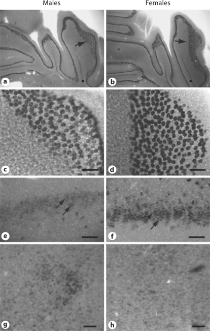
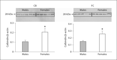
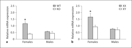
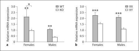
Most neurobehavioral diseases are sexually dimorphic in their incidence, and sex differences in the brain may be key for understanding and treating these diseases. Calbindin (Calb) D28K is used as a biomarker for the well-studied sexually dimorphic nucleus, a hypothalamic structure that is larger in males than in females. In the current study weanling C56BL/6J mice were used to examine sex differences in the Calb protein and message focusing on regions outside of the hypothalamus. A robust sex difference was found in Calb in the frontal cortex (FC) and cerebellum (CB; specifically in Purkinje cells); mRNA and protein were higher in females than in males. Using 2 mouse lines, i.e. one with a complete deletion of estrogen receptor alpha (ERα) and the other with uncoupled gonads and sex chromosomes, we probed the mechanisms that underlie sexual dimorphisms. In the FC, deletion of ERα reduced Calb1 mRNA in females compared to males. In addition, females with XY sex chromosomes had levels of Calb1 equal to those of males. Thus, both ERα and the sex chromosome complement regulate Calb1 in the FC. In the CB, ERα knockout mice of both sexes had reduced Calb1 mRNA, yet sex differences were retained. However, the sex chromosome complement, regardless of gonadal sex, dictated Calb1 mRNA levels. Mice with XX chromosomes had significantly greater Calb1 than did XY mice. This is the first study demonstrating that sex chromosome genes are a driving force producing sex differences in the CB and FC, which are neuoranatomical regions involved in many normal functions and in neurobehavioral diseases.
Copyright © 2011 S. Karger AG, Basel.
Figures
References
-
- Jazin E, Cahill L. Sex differences in molecular neuroscience: From fruit flies to humans. Nat Rev Neurosci. 2010;11:9–17. - PubMed
-
- Cohen-Bendahan CC, van de Beek C, Berenbaum SA. Prenatal sex hormone effects on child and adult sex-typed behavior: methods and findings. Neurosci Biobehav Rev. 2005;29:353–384. - PubMed
-
- Cooke B, Hegstrom CD, Villeneuve LS, Breedlove SM. Sexual differentiation of the vertebrate brain: principles and mechanisms. Front Neuroendocrinol. 1998;19:323–362. - PubMed
