

Human Ecstasy use is associated with increased cortical excitability: an fMRI study
- PMID: 21326196
- PMCID: PMC3079831
- DOI: 10.1038/npp.2010.244
Human Ecstasy use is associated with increased cortical excitability: an fMRI study
Abstract
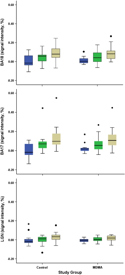
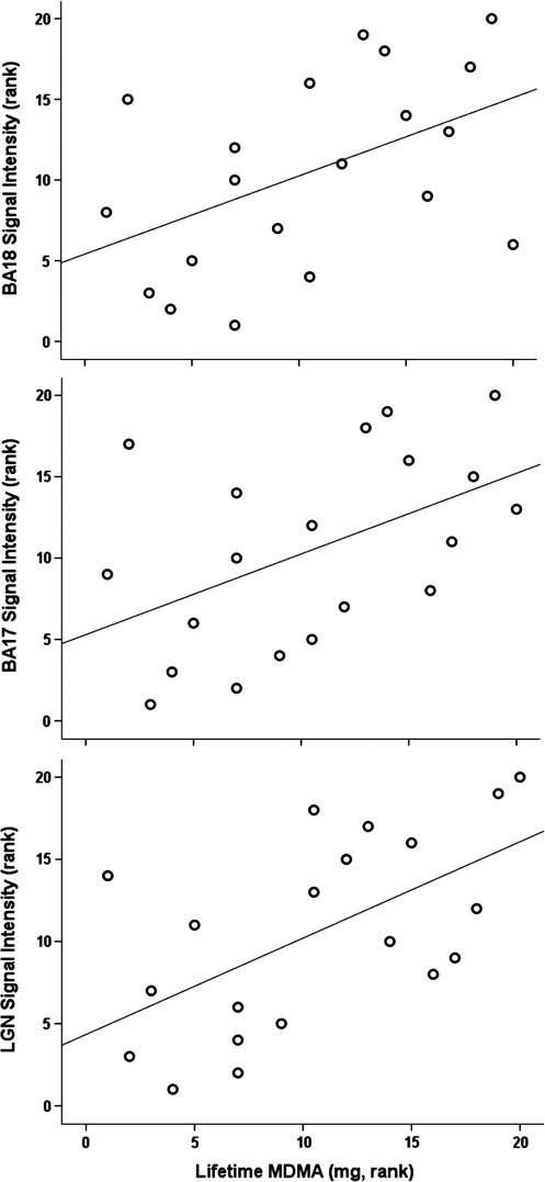
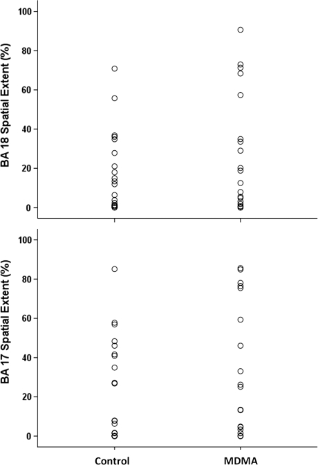
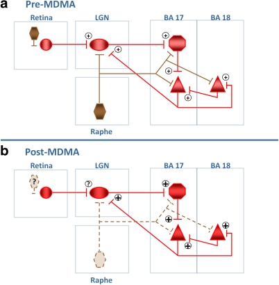
The serotonergic neurotoxin, 3,4-methylenedioxymethamphetamine (MDMA/Ecstasy), is a highly popular recreational drug. Human recreational MDMA users have neurocognitive and neuropsychiatric impairments, and human neuroimaging data are consistent with animal reports of serotonin neurotoxicity. However, functional neuroimaging studies have not found consistent effects of MDMA on brain neurophysiology in human users. Several lines of evidence suggest that studying MDMA effects in visual system might reveal the general cortical and subcortical neurophysiological consequences of MDMA use. We used 3 T functional magnetic resonance imaging during visual stimulation to compare visual system lateral geniculate nucleus (LGN) and Brodmann Area (BA) 17 and BA 18 activation in 20 long abstinent (479.95±580.65 days) MDMA users and 20 non-MDMA user controls. Lifetime quantity of MDMA use was strongly positively correlated with blood oxygenation level-dependent (BOLD) signal intensity in bilateral LGN (r(s)=0.59; p=0.007), BA 17 (r(s)=0.50; p=0.027), and BA 18 (r(s)=0.48; p=0.031), and with the spatial extent of activation in BA 17 (r(s)=0.059; p=0.007) and BA 18 (r(s)=0.55; p=0.013). There were no between-group differences in brain activation in any region, but the heaviest MDMA users showed a significantly greater spatial extent of activation than controls in BA 17 (p=0.031) and BA 18 (p=0.049). These results suggest that human recreational MDMA use may be associated with a long-lasting increase in cortical excitability, possibly through loss of serotonin input to cortical and subcortical regions. When considered in the context of previous results, cortical hyper-excitability may be a biomarker for MDMA-induced serotonin neurotoxicity.
Figures
 , low;
  , medium; and
  , high.
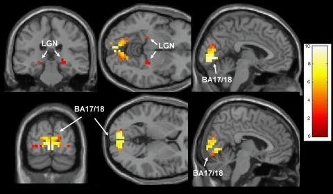

Comment in
-
Methodological weaknesses in non-randomized studies of ecstasy (MDMA) use: a cautionary note to readers and reviewers.Neuropsychopharmacology. 2012 Mar;37(4):1070-1; author reply 1072-3. doi: 10.1038/npp.2011.202. Neuropsychopharmacology. 2012. PMID: 22327903 Free PMC article. No abstract available.
References
-
- Bagdy G, Kecskemeti V, Riba P, Jakus R. Serotonin and epilepsy. J Neurochem. 2007;100:857–873. - PubMed
-
- Bankson MG, Cunningham KA. 3,4-Methylenedioxymethamphetamine (MDMA) as a unique model of serotonin receptor function and serotonin-dopamine interactions. J Pharmacol Exp Ther. 2001;297:846–852. - PubMed
-
- Bennett MR, Farnell L, Gibson WG. Origins of the BOLD changes due to synaptic activity at astrocytes abutting arteriolar smooth muscle. J Theor Biol. 2008;252:123–130. - PubMed
Publication types
MeSH terms
Substances
Grants and funding
LinkOut - more resources
Full Text Sources
Medical
