. 2011 Mar 16;30(6):1173-83.
doi: 10.1038/emboj.2011.14. Epub 2011 Feb 15.

The Arabidopsis peptide kiss of death is an inducer of programmed cell death

Affiliations

The Arabidopsis peptide kiss of death is an inducer of programmed cell death

Robert Blanvillain et al. EMBO J. .

Abstract

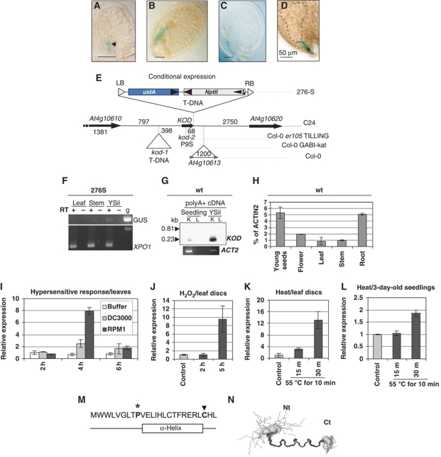
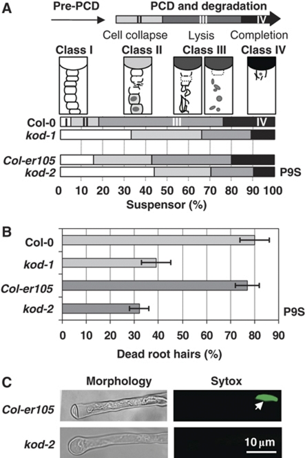
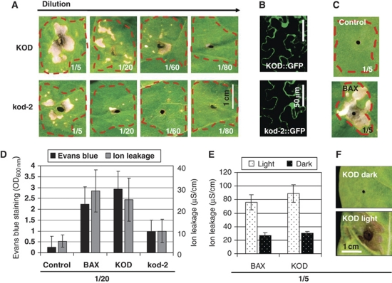
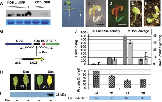
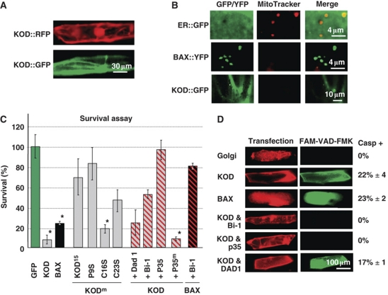
Programmed cell death (PCD) has a key role in defence and development of all multicellular organisms. In plants, there is a large gap in our knowledge of the molecular machinery involved at the various stages of PCD, especially the early steps. Here, we identify kiss of death (KOD) encoding a 25-amino-acid peptide that activates a PCD pathway in Arabidopsis thaliana. Two mutant alleles of KOD exhibited a reduced PCD of the suspensor, a single file of cells that support embryo development, and a reduced PCD of root hairs after a 55°C heat shock. KOD expression was found to be inducible by biotic and abiotic stresses. Furthermore, KOD expression was sufficient to cause death in leaves or seedlings and to activate caspase-like activities. In addition, KOD-induced PCD required light in leaves and was repressed by the PCD-suppressor genes AtBax inhibitor 1 and p35. KOD expression resulted in depolarization of the mitochondrial membrane, placing KOD above mitochondria dysfunction, an early step in plant PCD. A KOD∷GFP fusion, however, localized in the cytosol of cells and not mitochondria.


Conflict of interest statement

The authors declare that they have no conflict of interest.

Figures

Figure 1
Figure 1
Identification and expression profile of KOD. (AE) Trap line and KOD locus. GUS expression pattern in line 276S. Arrowhead shows the boundary between the suspensor and the embryo proper. Stages of embryo development (A) globular, (B) late globular, (C) heart, (D) torpedo. Bars=50 μm. (E) Locus and structure of the promoter trap T-DNA in 276S (ecotype C24). At4g10613: LINE retrotransposon in Col-0; kod-1: T-DNA position in the GABI-Kat line. P9S, non-synonymous substitution in kod-2 (TILLING). uidA, β-glucuronidase gene. DNA sizes are given in bp. (FL) Expression analysis of KOD. (F) RT–PCR analysis on tissues from line 276S using primer oexTi15 in KOD and oGUSj in the GUS gene. ‘GUS’ indicates the transcriptional fusion between KOD and the GUS sequence. XPO1, a ubiquitous gene (At5g17020) as a control. g, genomic DNA; YSil, young silique. (G) RT–PCR on polyA+ cDNA of WT, blotted and probed with a radiolabelled KOD ORF. K, Primer pair (oexTi15; oSUPR1) amplifying the KOD transcript (0.23 kb); L, primer pair (oTi05; oSUPR1) with oTi05 outside the transcript (0.81 kb). Expected sizes are shown; ACT2, Actin2 control. (H) KOD expression in Col-0 wild-type tissues using Taqman QRT–PCR. Actin2 is used as the reference gene. (I) KOD expression using Taqman QRT–PCR in WT leaves following infiltration with 10 mM MgCl2 (Buffer), P. syringae pv. tomato DC3000 wild-type (virulent) or expressing AvrRPM1 (avirulent). 18S is used as the reference gene. Each sample was measured in triplicate and is shown relative to expression at 2 h post-inoculation (hpi). (J) KOD expression using Taqman QRT–PCR in WT leaf discs floated on SDW (control) or 30 mM H2O2 for 2 and 5 h. 18S is used as the reference gene. Each sample was measured in triplicate and is shown relative to expression in the control. (K) KOD expression using Taqman QRT–PCR in WT leaf discs floated on SDW at RT (control) or 15 and 30 min after heat shock at 55°C for 10 min. 18S is used as the reference gene. Each sample was measured in triplicate and is shown relative to expression of control. (L) KOD expression using Taqman QRT–PCR in WT 3-day-old seedlings floated on SDW at RT (control) or 15 and 30 min after heat shock at 55°C for 10 min. 18S is used as the reference gene. Each sample was measured in triplicate and is shown relative to expression of control. (M) Amino-acid sequence of KOD and position of α-helix, see Supplementary Figure S1. Star and arrow highlight 2 aa required for full functionality of KOD, see Figure 5C. (N) Overlay of 22 calculated structures from NMR spectra. Nt, N-terminus; Ct, C-terminus. Residues 9–21-fold into an α-helix, whereas the N-terminal region is disordered.
Figure 2
Figure 2
Phenotype in Arabidopsis KOD mutant lines. Mutant lines are kod-1, T-DNA knockdown (GABI line); kod-2, P9S point mutation in Col-er105 background (TILLING line). (A) Stacked column chart with the percentage of suspensors in classes I–IV for 100 embryos of each genotype. Seeds containing embryos at the bent-cotyledon stage were clarified using Hoyers solution and observed under DIC. Each suspensor was assigned to one of the four stages (classes I–IV) of suspensor elimination described in Supplementary Figure S2. (B) Reduced root hair PCD in 3-day-old seedlings of the two mutant backgrounds. PCD was induced using 10 min at 55°C. Cell death was scored 6 h after treatment using plasma membrane retraction and sytox green positive nucleus as criteria. Untreated seedlings of all backgrounds had around 10% dead root hairs. Errors bars are 2 × s.e. of triplicates of five seedlings (>100 root hairs total) each. (C) Most common root hair morphology in wild-type Col-er105 (dead root hair) or kod-2 (live root hair). (Left panel) White light microscopy. (Right panel) Fluorescence microscopy of one root hair incubated with sytox green using FITC filter. White arrow points at a sytox-positive nucleus, indicative of a loss of plasma membrane permeability.
Figure 3
Figure 3
KOD expression in N. tabacum leaves induces death. (A) Agrobacterium-mediated transfection of tobacco leaves using 35S∷KOD∷GFP and 35S∷kod2∷GFP. OD600 for each sample was adjusted to 1 and further diluted to create the series, that is 1/5=OD 0.2. Phenotypes were seen 3 dpi. (B) Fluorescent confocal microscopy confirming GFP expression in the infiltrated samples, scale bar=50 μm. (C) 35S∷GFP control and 35S∷BAX∷YFP infiltrations. (D) Quantification of cell death for 1/20 dilutions, using leaf discs for both ion leakage and Evans blue in triplicate ±2 × s.e. (E) Tobacco leaves were infiltrated with the OD 0.2 (1/5) dilution of KOD∷GFP, BAX∷YFP and GFP. Leaves either kept in 8 h light/16 h dark or wrapped in tin foil to exclude light. Ion leakage was taken at 4 dpi; measurements are minus the average reading for the GFP control. Values are taken in triplicate, ±2 × s.e. (F) Photos (4 dpi) of KOD infiltrations.
Figure 4
Figure 4
KOD overexpression in Arabidopsis correlates with PCD. (A) Western blot analysis using anti-GFP antibody on 35S∷KOD∷GFP and 35S∷KOD15∷GFP Arabidopsis lines. T1 plants were grouped according to their GFP signal intensity. Equal loading was controlled with Coomassie blue staining. (BF) F1 35S∷KOD∷GFP plants. (B) Unaffected seedling (left) compared with most affected kanamycinR seedlings (top right) and to kanamycinS seedlings (bottom right). Magnified view in bright field of an affected kanamycinR seedling (C) and under blue light excitation (D), scale bars=1 mm. (G) Cartoon of the pOp-LhGR/dex 2-component inducible system for conditional expression. (H) Leaves after 3 days of induction in continuous light. Individual leaves were kept in 1.5 ml Eppendorf tubes with their petioles submerged in 30 μM dex. (I) Western using anti-GFP on protein extracted from wt leaves (±dex) and high expresser line 23 (±dex), after 3 days with 30 μM dex. (J) (Top panel) Quantification of cell death using ion leakage and caspase3-like activity in three inducible KOD lines after 3 days of induction. (Middle panel) Quantification of total protein loss measured with the Bradford assay (7 days of induction), values from triplicate with ±2 × s.e. (Bottom panel) Fold dex induction of expression using a GUS fluorometric assay.
Figure 5
Figure 5
KOD-induced PCD pathway. (A) Subcellular localization of KOD∷GFP and KOD∷RFP in onion epidermal cells 15 h post-bombardment (pb). (B) Onion cells 24 h pb, incubated in 100 nM of MitoTracker Red for 5 min. Absence of MitoTracker signal indicates loss of ψmit. ER∷GFP, ER-targeted GFP. (C) Survival assay in onion cells quantified as the percentage of cells with no pH shift (fluorescent cells). KODm are P9S, C16S and C23S single missense mutations. AtBi-1, p35 and Dad1 are used as suppressors of PCD; p35m is the null mutant allele D87A of p35. All suppressor plasmids were co-bombarded in a two-fold molar excess over KOD or BAX. Values from triplicates ±2 × s.e. (D) In situ fluorescent caspase assay in transfected onion cells. Onion cells incubated with FAM-VAD-FMK inhibitor at 30 h pb. A small number of cells with caspase activity was observed in the negative control (golgi∷RFP) and was subtracted from all other samples. Values from triplicates with ±2 × s.e.

References

    1. Allen LJ, MacGregor KB, Koop RS, Bruce DH, Karner J, Bown AW (1999) The relationship between photosynthesis and a mastoparan-induced hypersensitive response in isolated mesophyll cells. Plant Physiol 119: 1233–1242 - PMC - PubMed
    1. Bechtold N, Pelletier G (1998) In planta Agrobacterium-mediated transformation of adult Arabidopsis thaliana plants by vacuum infiltration. Methods Mol Biol 82: 259–266 - PubMed
    1. Bonneau L, Ge Y, Drury GE, Gallois P (2008) What happened to plant caspases? J Exp Bot 59: 491–499 - PubMed
    1. Bozhkov P, Filonova L, Suarez M (2005) Programmed cell death in plant embryogenesis. Curr Topics Dev Biol 67: 135–179 - PubMed
    1. Bozhkov PV, Filonova LH, Suarez MF, Helmersson A, Smertenko AP, Zhivotovsky B, von Arnold S (2003) VEIDase is a principal caspase-like activity involved in plant programmed cell death and essential for embryonic pattern formation. Cell Death Differ 11: 175–182 - PubMed

Publication types