. 2011 Jul;12(5):360-9.
doi: 10.1038/gene.2011.4. Epub 2011 Feb 17.

Interferon-inducible gene 202b controls CD8(+) T cell-mediated suppression in anti-DNA Ig peptide-treated (NZB × NZW) F1 lupus mice

Affiliations
Free PMC article

Interferon-inducible gene 202b controls CD8(+) T cell-mediated suppression in anti-DNA Ig peptide-treated (NZB × NZW) F1 lupus mice

R Dinesh et al. Genes Immun. 2011 Jul.
Free PMC article

Abstract

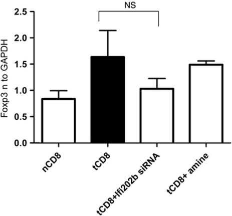
Administration of an artificial peptide (pConsensus) based on anti-DNA IgG sequences that contain major histocompatibility complex class I and class II T-cell determinants, induces immune tolerance in NZB/NZW F1 female (BWF1) mice. To understand the molecular basis of CD8(+) Ti-mediated suppression, we previously performed microarray analysis to identify genes that were differentially expressed following tolerance induction with pCons. CD8(+) T cells from mice tolerized with pCons showed more than two-fold increase in Ifi202b mRNA, an interferon inducible gene, versus cells from untolerized mice. Ifi202b expression increased through weeks 1-4 after tolerization and then decreased, reapproaching baseline levels at 6 weeks. In vitro polyclonal activation of tolerized CD8(+) T cells significantly increased Ifi202b mRNA expression. Importantly, silencing of Ifi202b abrogated the suppressive capacity of CD8(+) Ti cells. This was associated with decreased expression of Foxp3, and decreased gene and protein expression of transforming growth factor (TGF)β and interleukin-2 (IL-2), but not of interferon (IFN)-γ, IL-10, or IL-17. Silencing of another IFN-induced gene upregulated in tolerized CD8(+) T cells, IFNAR1, had no effect on the ability of CD8(+) T cells to suppress autoantibody production. Our findings indicate a potential role for Ifi202b in the suppressive capacity of peptide-induced regulatory CD8(+) Ti cells through effects on the expression of Foxp3 and the synthesis of TGFβ.


Figures

Figure 1
Figure 1
(a) Ifi202b mRNA expression is dynamic after pCons tolerization in BWF1 mice. Saline or negative control peptide-treated naïve CD8+ T cells and pCons-tolerized CD8+ T cells (1–2 × 10⁶ cells) were isolated from BWF1 mice splenocytes at different times (2, 4 and 6 weeks after pCons treatment). RNA was isolated and real-time PCR was performed with murine Ifi202b primers and probes. An Ifi202b standard curve was created and the relative input amount was calculated. GAPDH was used as a housekeeping gene. Results are shown from three independent experiments from 3 to 5 mice in each group. Horizontal lines indicate means. **P<0.001, *P<0.03, statistically significant. (bd). Expression of Ifi202b changes spontaneously in spleen cells of BWF1 mice with age and development of disease, starts to decrease at 50 weeks of age in CD4+ T cells, and exhibits little to no change in CD8+ T cells. In contrast, in B cells, Ifi202b increases spontaneously until the 50th week of age. CD4+ T cells, CD8+ T cells and B cells were isolated from spleens of 3–5 unmanipulated BWF1 females at different ages (6, 20, and 50 weeks of age). RNA was isolated and real-time PCR was performed. Data shown are from three experiments. (eg). Ifi202b protein was increased after pCons tolerance. Western blot analyses were performed with Ifi202b-specific antibody using cell lysates from CD8+ T cells, B cells and splenocytes from naïve and pCons-treated mice (3–5 mice in each group). The β-actin was used for normalization purposes in the densitometry analyses.
Figure 2
Figure 2
Tolerized CD8+ T cells suppress autoantibody production at 6 weeks after pCons treatment. CD8+ T cells were isolated 6 weeks after pCons treatment and cells were cultured with naïve CD4 T cells and B cells (Spleens were pooled from 3 to 4 mice per group). After 3–4 days, the culture supernatants were collected and anti-DNA was measured by ELISA. Tolerized CD8+ Ti suppress anti-DNA production significantly (column 3 compare with column 2, P<0.04, paired one-tailed test); that suppression was lost after silencing of Ifi202b (column 4), *P<0.02 by paired two-tailed test, nB+nCD4+tCD8 vs nB+nCD4+tCD8+Ifi202b siRNA. Each dot represents data from each experimental replicate and the horizontal bar denotes means. Data shown are individual results from four independent experiments.
Figure 3
Figure 3
(a) Silencing with siRNA of Ifi202b abrogates suppression of anti-DNA induced by CD8+ Ti cells. CD8+ T cells were isolated from pCons-treated mice 1–2 weeks after pCons tolerization and the cells were transfected with either Ifi202b siRNA, IFNAR1 siRNA, or control siRNA, as described in the Materials and methods section. Then, non-transfected and transfected CD8+ T cells (1 × 10⁵) were cultured with CD4+ T cells (1 × 10⁵) and B cells (1 × 10⁴) from naïve BWF1 mice. After 4–5 days, culture supernatants were collected and measured for anti-DNA IgG with ELISA. Tolerized CD8=tCD8; Naïve CD8=nCD8. Columns: a, nCD4; b, nB; c, nCD4+ nB; d, nCD4+nB+tCD8; e, nCD4+nB+tCD8+Ifi202b siRNA; f, nCD4+nB+tCD8+IFNAR1siRNA; g, nCD4+nB+tCD8+si port Amine; h, nCD4+nB+tCD8+GAPDH siRNA; i, nCD4+nB+tCD8+GAPDH-negative siRNA or scrambled siRNA with no homology to mouse, rat or human genomes. Data shown are from three to four independent experiments. (b) Silencing of Ifi202b reduces Ifi202b protein. Tolerized CD8+ T cells (2–3 × 10⁶cells) were isolated and cultured with and without siRNA of Ifi202b (100, 50 n, scrambled siRNA (100 n)) for 48–72 h. Cell lysates were prepared and western blot was performed with Ifi202b and β-actin antibody as described in Materials and methods. Densitometry was performed (***P<0.0004 tCD8 vs tCD8+IFI202b siRNA 100 n. *P<0.011 tCD8 vs tCD8+IFI202b siRNA 50 n). Densitometry data shown are combined from five experiments.
Figure 4
Figure 4
Silencing of Ifi202b reduces Foxp3 expression in tolerized CD8+ T cells. Naïve and tolerized CD8+ T cells from 3 to 5 in each group were isolated and cultured as shown previously with Ifi202b siRNA and scrambled siRNA. RNA was isolated and equal amounts (100 ng) of RNA were used for real-time PCR with Foxp3 primers and probes (TaqMan—Applied Biosystems). Values were normalized to GAPDH. Paired one-tailed or two-tailed test was used. P<0.05 were considered significant. Data shown are from three experiments.
Figure 5
Figure 5
(a) Silencing of Ifi202b in tolerized CD8+ T cells decreases IL-2 protein secretion. Naïve and tolerized CD8+ T cells (1 × 10⁵) were cultured as shown previously with Ifi202b siRNA and scrambled siRNA. Culture supernatant was obtained and IL-2 secreted protein was measured by ELISA as per manufacturer's instructions (Biolegend Inc.). An IL-2 standard curve was created and unknown values were determined. Data shown are from two to four experiments. Error bars indicate s.e.m. Paired one or two-tailed t-test was used. *P<0.01 considered as significant. (b) Silencing of Ifi202b in tolerized CD8+ T cells decreases IFN-γ protein secretion. IFN-γ secreted protein was measured from the above culture using an IFN-γ mouse ELISA kit from BD Biosciences (San Diego, CA, USA) as per manufacturer's instructions. An IFN-γ standard curve was created and values were analyzed. Data shown are from two experiments. (c) Silencing of Ifi202b in tolerized CD8+ T cells does not change IL-10 protein secretion. IL-10 protein was measured from the above culture by ELISA as per manufacturer's instructions. Data shown are from two experiments. (d) Tolerized CD8+ T cells have reduced IL-17 secretion and Ifi202b silencing has no effect on IL-17. IL-17 was measured from the above culture supernatant using an ELISA kit from Biolegend as per manufacturer's instructions. An IL-17 standard curve was created and values were analyzed. Data shown are from three experiments. *P<0.03 considered as significant.
Figure 6
Figure 6
Silencing of Ifi202b in tolerized CD8+ T cells decreases TGFβ expression. (a) Tolerized CD8+ T cells (2–3 × 10⁶cells) were isolated and cultured with and without the siRNA of Ifi202b or IFNAR1 (50 n) for 48–72 h. RNA was isolated from these cultures and real-time PCR was performed. Silencing of Ifi202b in tolerized CD8+ T cells decreased TGFβ expression (*P<0.04 nCD8 vs tol CD8, **P<0.003 tCD8 vs tCD8+Ifi202b si RNA, paired two-tailed t-test). Error bars indicate s.e.m. Each dot represents data from each experimental replicate and the horizontal bar denotes means. Data shown are from two independent experiments performed. (b, c) IFI202b silencing reduced TGFβ mRNA and protein expression. TGFβ was measured from culture supernatant from naïve and tolerized CD8+ T cells with and without siRNA treatment. Each sample was run in duplicate and the values were determined from the standard curve of TGFβ. Silencing of Ifi202b significantly reduced TGFβ, *P<0.018, paired two-tailed t-test. Error bars denote s.e.m. Compare tolerized CD8+ T cells vs tolerized CD8+ T cells+Ifi202b siRNA. Data shown are from five experiments. (c) TGFβ mRNA was determined by real-time PCR as described previously. (d) Immunoblots were assayed from cell lysates prepared from splenocytes with and without siRNA treatment as above. Equal amount of protein (10–15 μg) was loaded in each well. Gels were transferred to a polyvinylidene fluoride (PVDF) membrane and immunoblotted with TGFβ and β-actin-specific antibodies. Lane 1—naïve splenocytes, lane 2—tolerized splenocytes, lane 3—tolerized splenocytes + Ifi202b siRNA, lane 4-tolerized splenocytes + scrambled siRNA. (e) Densitometry analysis of mature TGFβ normalized to β-actin. P<0.01 by paired two-tailed t-test. (f) Smad 2 and smad 3 mRNA expressions were determined by real-time PCR. Error bars denote s.e.m. Values were normalized to GAPDH.

Comment in

Similar articles

Cited by

References

    1. Kotzin BL. Systemic lupus erythematosus. Cell. 1996;85:303–306. - PubMed
    1. La Cava A, Ebling FM, Hahn BH. Ig-reactive CD4+CD25+ T cells from tolerized (New Zealand Black x New Zealand White) F1 mice suppress in vitro production of antibodies to DNA. J Immunol. 2004;173:3542–3548. - PubMed
    1. Earle K E, Tang Q, Zhou X, Liu W, Zhu S, Bonyhadi ML, et al. In vitro expanded human CD4+CD25+ regulatory T cells suppress effector T cell proliferation. Clin Immunol. 2005;115:3–9. - PMC - PubMed
    1. Singh RP, La Cava A, Wong M, Ebling F, Hahn BH. CD8+ T cell-mediated suppression of autoimmunity in a murine lupus model of peptide-induced immune tolerance depends on Foxp3 expression. J Immunol. 2007;178:7649–7657. - PubMed
    1. Suzuki M, Konya C, Goronzy JJ, Weyand CM. Inhibitory CD8+ T cells in autoimmune disease. Hum Immunol. 2008;69:781–789. - PMC - PubMed

Publication types

MeSH terms