

doi: 10.1055/s-0028-1085921.
Imaging and percutaneous treatment of vascular anomalies
Affiliations
- PMID: 21326512
- PMCID: PMC3036444
- DOI: 10.1055/s-0028-1085921
Item in Clipboard
Imaging and percutaneous treatment of vascular anomalies
Semin Intervent Radiol.
2008 Sep.
Abstract
Vascular anomalies are an extensive group of malformations of the arterial, venous, and lymphatic systems, either in isolation or, more often, in combination. Although mostly congenital, they can occasionally be acquired as well. They present a challenge both for workup and therapy. This article attempts to describe some of their main anomalies, their workup, and their therapies, with the goal of increasing the comfort level of endovascular therapists.
Keywords: Vascular anomalies; embolization; hemangiomas; malformations; percutaneous endovascular therapy.
Figures
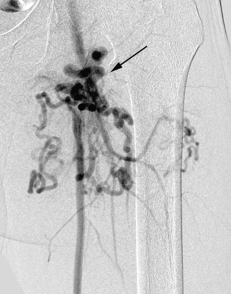
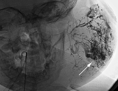
Digital subtraction angiography of a thigh arteriovenous malformation shows a nidus (arrow) with multiple feeders from the profunda femoris and superficial femoral artery.
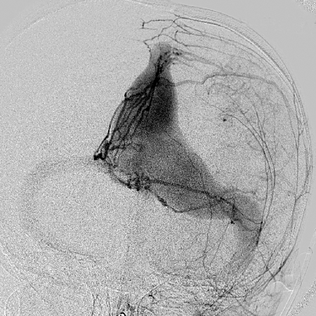
Left external carotid angiogram showing direct shunting from arterial branches to venous sinus without presence of a nidus is suggestive of a dural arteriovenous fistula.
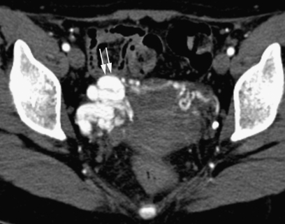
Axial contrast-enhanced computed tomography of the pelvis shows a nidus with dilated vessels in the right adnexa (double arrow).
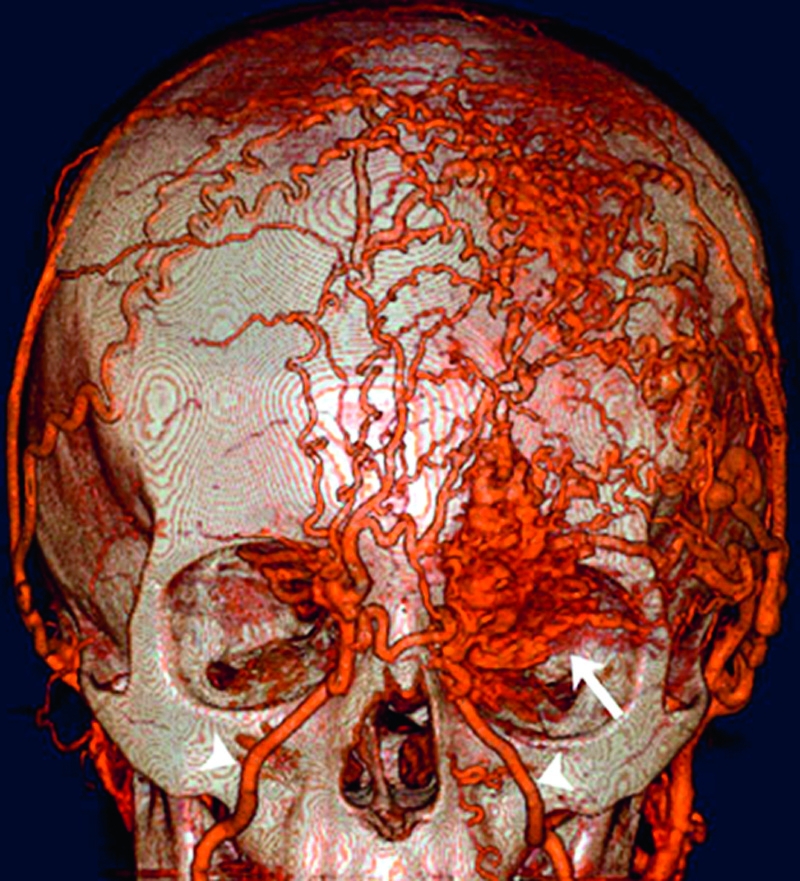
Three-dimensional computed tomography angiography of face shows an arteriovenous malformation involving the left orbit. Feeding vessels (arrowheads) and nidus (arrow) are clearly depicted.
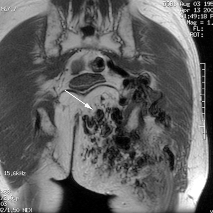
Coronal T2-weighted magnetic resonance imaging of gluteal region showing a large arteriovenous malformation with hypointense areas (arrow) is suggestive of flow voids, indicating high flow.
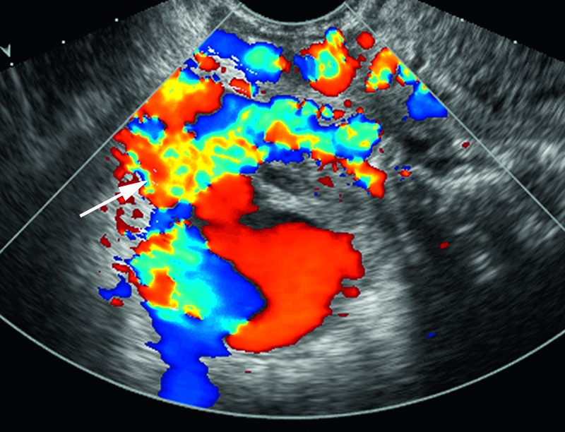
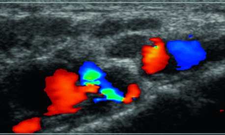
Color Doppler ultrasound of the pelvis (right adnexa) showing a large hypervascular area with aliasing in the high-flow areas (arrow) is suggestive of an arteriovenous malformation.
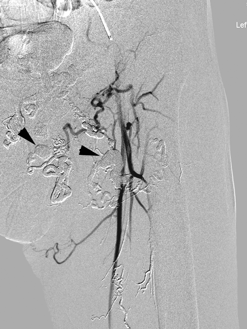
Postembolization angiogram of thigh arteriovenous malformation (AVM) shows nonfilling of the AVM with Onyx and glue casts seen (arrowheads).
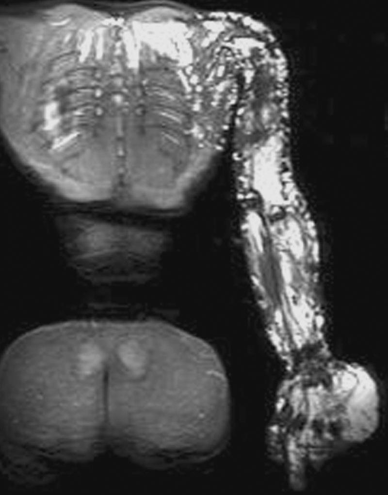
Magnetic resonance T2 fat-saturated image shows an extensive upper extremity venous malformation that extends into the chest wall. Note the involvement of the underlying muscles in the upper extremity.
Color Doppler ultrasound of a venous malformation in the dorsum of the foot. Note lack of aliasing, indicating slow flow in contrast to arteriovenous malformations.
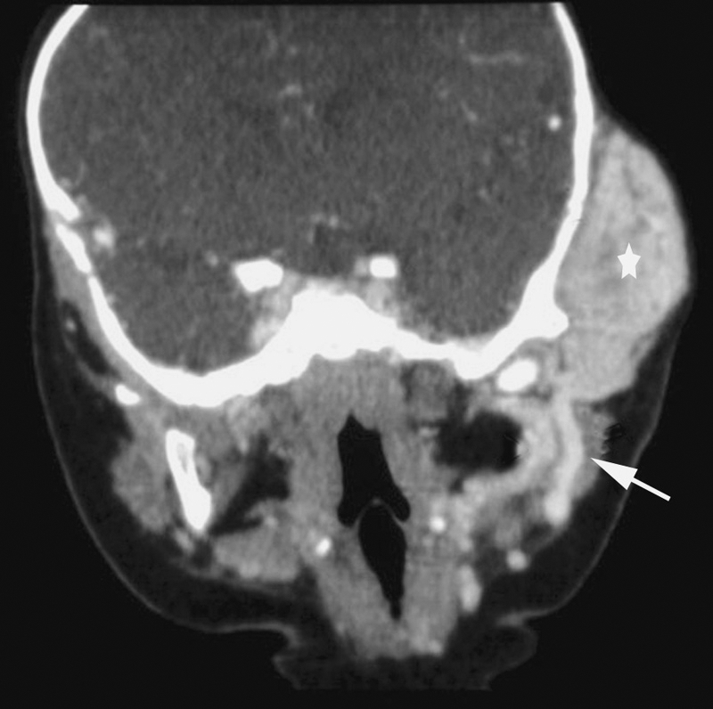
Coronal contrast enhanced computed tomography of a noninvoluting congenital hemangioma involving the left lateral cheek and temporal region shows an intensely enhancing mass (*) with a feeder vessel (arrow).
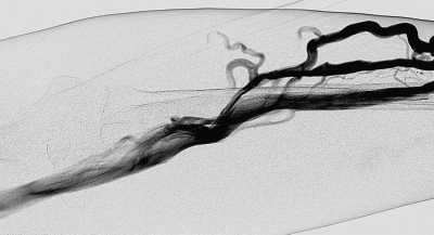
Venogram in a patient with Parks-Weber syndrome shows anomalous deep venous anatomy. This was in addition to the high-flow lesions (not shown).
Spot image during angiogram for preoperative embolization shows a large facial venous malformation with liquid embolic material (arrow) in it.
Venogram during sclerotherapy of a venous malformation shows venous channels and lakes.
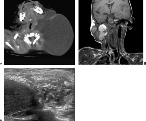
(A) Contrast-enhanced axial computed tomography through the neck shows a large nonenhancing cystic mass consistent with cystic hygroma. (B) Mixed macro- and microcystic lymphatic malformation. T1-weighted coronal shows the macrocystic portion as high signal, with the superficial microcystic portion minimally hyperintense to muscle. (C) Ultrasound shows the more homogeneous macrocystic portion containing swirling proteinatious fluid, with superficial microcystic region.
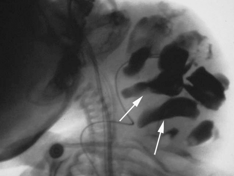
Lateral spot image during injection sclerotherapy shows the sclerosant opacified with contrast outlining the different cystic compartments (arrows).
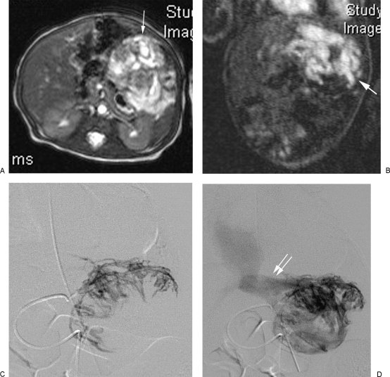
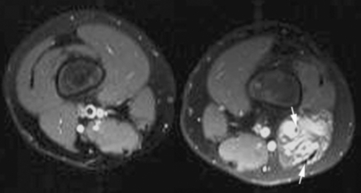
Axial (A) and coronal (B) T1 fat-saturated postcontrast images. A large enhancing mass is seen in the left lobe of the liver (arrow). Selective left hepatic angiogram shows classical peripheral filling (C) of this mass, and a subsequent image (D) shows early filling of the venous outflow in the form of the dilated left hepatic vein (double arrows).
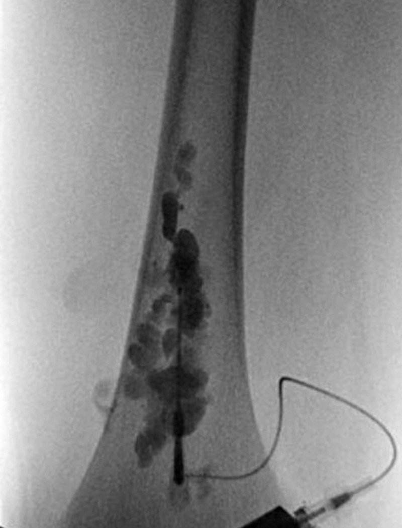
Axial postcontrast fat-saturated T1-weighted image through the thigh shows an enhancing lesion in the left lateral thigh with phleboliths (arrows) in it.
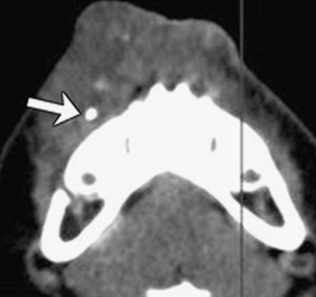
Noncontrast axial computed tomography of the mandible shows a soft tissue mass in the superficial tissues with a phlebolith (arrow) in it suggestive of a hemangioma.
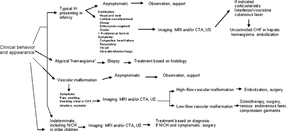
Flowchart provides a sample protocol for managing vascular anomalies, including multidisciplinary workup and treatment for vascular anomalies. IH, infantile hemangioma; MRI, magnetic resonance imaging; CTA, computed tomography angiography; US, ultrasound; CHF, congenital hepatic fibrosis; NICH, Noninvoluting congenital hemangioma.
Similar articles
-
Peripheral Vascular Anomalies - Essentials in Periinterventional Imaging.Rofo. 2020 Feb;192(2):150-162. doi: 10.1055/a-0998-4300. Epub 2019 Oct 17. Rofo. 2020. PMID: 31622988 English.
-
[Interdisciplinary concept for classification and treatment of vascular anomalies in the head and neck].Mund Kiefer Gesichtschir. 2002 Nov;6(6):402-9. doi: 10.1007/s10006-002-0418-z. Epub 2002 Aug 9. Mund Kiefer Gesichtschir. 2002. PMID: 12447652 German.
-
Congenital vascular malformations: when and how to treat them.Semin Vasc Surg. 2002 Mar;15(1):65-71. doi: 10.1053/svas.2002.30450. Semin Vasc Surg. 2002. PMID: 11840428 Review.
-
[Extracranial vascular anomalies (hemangiomas and vascular malformations) in children and adolescents--diagnosis, clinic, and therapy].Laryngorhinootologie. 2014 Mar;93 Suppl 1:S185-202. doi: 10.1055/s-0033-1363216. Epub 2014 Apr 7. Laryngorhinootologie. 2014. PMID: 24710783 Review. German.
-
Endovascular methods for the treatment of vascular anomalies.Neuroimaging Clin N Am. 2013 Nov;23(4):703-28. doi: 10.1016/j.nic.2013.03.016. Epub 2013 May 30. Neuroimaging Clin N Am. 2013. PMID: 24156860 Review.
Cited by
-
Endovascular balloon-assisted liquid embolisation of soft tissue vascular malformations: technical feasibility and safety.CVIR Endovasc. 2021 Jun 8;4(1):49. doi: 10.1186/s42155-021-00236-4. CVIR Endovasc. 2021. PMID: 34101056 Free PMC article.
-
Pediatric Vascular Anomalies: A Clinical and Radiological Perspective.Indian J Radiol Imaging. 2023 Sep 16;34(1):103-127. doi: 10.1055/s-0043-1774391. eCollection 2024 Jan. Indian J Radiol Imaging. 2023. PMID: 38106867 Free PMC article. Review.
-
A Step-by-Step Practical Approach to Imaging Diagnosis and Interventional Radiologic Therapy in Vascular Malformations.Semin Intervent Radiol. 2010 Jun;27(2):209-31. doi: 10.1055/s-0030-1253521. Semin Intervent Radiol. 2010. PMID: 21629410 Free PMC article.
References
-
- Mulliken J B, Glowacki J. Hemangiomas and vascular malformations in infants and children: a classification based on endothelial characteristics. Plast Reconstr Surg. 1982;69(3):412–422. - PubMed
-
- JK W, TS L, CT R, et al. For hemangiomas refractory to first line corticosteroid therapy, interferon and vincristine therapy has been described. Wellington, New Zealand: 2004. In, International Society for the Study of Vascular Anomalies.
-
- Chang M W. Updated classification of hemangiomas and other vascular anomalies. Lymphat Res Biol. 2003;1(4):259–265. - PubMed
-
- Upton J, Coombs C J, Mulliken J B, Burrows P E, Pap S. Vascular malformations of the upper limb: a review of 270 patients. J Hand Surg [Am] 1999;24(5):1019–1035. - PubMed
-
- Cho S K, Do Y S, Shin S W, Kim D I, et al. Arteriovenous malformations of the body and extremities: analysis of therapeutic outcomes and approaches according to a modified angiographic classification. J Endovasc Ther. 2006;13:527–538. - PubMed
