. 2008 Sep;25(3):293-309.
doi: 10.1055/s-0028-1085929.

Embolization in the head and neck

Affiliations

Embolization in the head and neck

Daniel Cooke et al. Semin Intervent Radiol. 2008 Sep.

Abstract

In this article, we review current practices in therapeutic embolization of the head and neck. Major applications including vascular malformations, highly vascular tumors, trauma, and other sources of hemorrhage are discussed. We emphasize the importance of a thorough knowledge of head and neck vascular anatomy, especially of potential connections to critical territories not intended for embolization. The choice of embolic agent and its effect on safety and efficacy of treatment are presented.

Keywords: Intervention; bleeding; head and neck; tumor; vascular malformation.


Figures

Figure 1
Figure 1
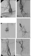
(A) Large right cervical arteriovenous malformation is supplied by right thyrocervical artery, costocervical artery, and right vertebral artery branches. Note dysplastic venous drainage. (B) Supply from the right vertebral artery with anastomosis between the ascending cervical and right vertebral artery is seen. (C) Selective injection of the thyrocervical feeders shows intranidal arteriovenous fistulas (AVFs). (D) Coil embolization of the AVFs with electrolytically detachable coils is shown. (E) Coil embolization of communication between the vertebral and ascending cervical branches is shown. (F) Lateral view shows coil and n-butyl-2 cyanoacrylate cast of the nidus following embolization. (G) Anteroposterior (AP) angiogram shows post-embolization of right subclavian artery. (H) At 6-month follow-up, right subclavian angiogram, AP view, shows minimal residual nidus.
Figure 2
Figure 2
(A) Left common carotid artery angiogram shows large left parietal scalp arteriovenous malformation (AVM) supplied by superficial temporal and occipital branches of the left external carotid artery (ECA). (B) Unsubtracted image of left CCA injection as described in 2A. Selective injection performed of feeding artery from the left superficial temporal artery before Onyx injection. (C) Onyx cast of the AVM is shown. (D) Angiogram shows postembolization of left ECA. (E) Delayed postembolization ECA angiogram shows complete obliteration.
Figure 3
Figure 3
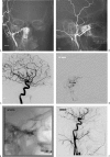
(A) Right cervical paraganglioma is supplied by branches of ascending pharyngeal and superior thyroid branches of external carotid artery (ECA) and right internal carotid artery (ICA), splaying the carotid bifurcation (anteroposterior [AP] view). (B) Right cervical paraganglioma is supplied by branches of ascending pharyngeal and superior thyroid branches of ECA and right ICA, splaying the carotid bifurcation (lateral view). (C) Selective injections of ascending pharyngeal artery were performed after occluding neuromeningeal trunk with Gelfoam torpedo for protection of cranial nerve supply. (D) Selective superior thyroid injections performed before embolization show tumor blush. (E) AP view of postembolization of right common carotid artery (CCA) angiogram following embolization with 150–250-μm polyvinyl alcohol (PVA) particles is seen. (F) Lateral view of postembolization of right CCA angiogram following embolization with 150–250-μm PVA particles is seen.
Figure 4
Figure 4
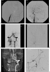
(A) Juvenile right posterior nasal angiofibroma is supplied by sphenopalatine and pterygopalatine branches of internal maxillary artery (IMA) (anteroposterior [AP] view). (B) Juvenile right posterior nasal angiofibroma is supplied by sphenopalatine and pterygopalatine branches of IMA (lateral view). (C) Vidian artery supply is shared with the right internal carotid artery. (D) Selective pterygopalatine is injected before Onyx embolization. (E) Onyx cast of tumor is shown. (F) Postembolization of right common carotid artery angiogram shows marked reduction of tumor blush.
Figure 5
Figure 5
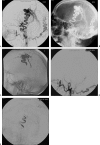
(A) Lateral view of right external carotid artery (ECA) shows supply to the nasal region by branches of the internal maxillary artery (IMA) and facial artery. (B) Lateral view of right internal carotid artery (ICA) shows that normal chorioretinal blush from the ophthalmic artery has a normal origin from the ICA. Some nasal blush from the ethmoidal branches of the ophthalmic is also seen. (C) Lateral view of right ECA injection after embolization of the IMA reveals prominent supply to the nasal region via the facial artery, signifying importance of also embolizing the facial artery. (D) Anteroposterior view of left ECA injection shows filling of right IMA branches through collaterals, signifying importance of bilateral embolization.
Figure 6
Figure 6
(A) Right vertebral angiogram shows complete occlusion of the artery at the level of C3-C4 following gunshot injury. (B) Left common carotid artery angiogram shows patent posterior communicating artery with opacification of distal right vertebral artery. (C) Left vertebral angiogram shows retrograde flow into distal right vertebral venous fistula. There are no opacifications of distal basilar artery. (D) Microcatheter is placed at site of fistula from left vertebral basilar artery into right vertebral artery. (E) Coil mass is seen in the fistula site with antegrade flow into basilar artery through left vertebral injection. (F) Right subclavian angiogram shows deep cervical collaterals opacifying the distal right vertebral artery after obliteration of fistula.

References

    1. Morris P P. Practical Neuroangiography. 2nd ed. Philadelphia: Lippincott, Williams, & Wilkins; 2006.
    1. Byrne J. Interventional Neuroradiology. Oxford, UK: Oxford University Press; 2002.
    1. Osborn A. Diagnostic Angiography. 2nd ed. Philadelphia: Lippincott, Williams, & Wilkins; 1999.
    1. Bouthillier A, Loveren H van, Keller J. Segments of the internal carotid artery: a new classification. Neurosurgery. 1996;38(3):425–432. discussion 432–433. - PubMed
    1. Lapresle J, Lasjaunias P. Cranial nerve ischaemic arterial syndromes. A review. Brain. 1986;109(Pt 1):207–216. - PubMed