

The evaluation of lower-extremity ulcers
- PMID: 21326538
- PMCID: PMC3036466
- DOI: 10.1055/s-0029-1242204
The evaluation of lower-extremity ulcers
Abstract
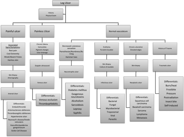
Lower-limb ulceration is prevalent in Western countries. There are many different types of ulcers with several causes. The most prevalent are those due to vascular disease, of which venous is the most common, accounting for over two-thirds of all types of ulcers. There are also many other causes for ulceration such as malignancy, infections, and skin, drug-induced, and autoimmune diseases. The ulcers have different characteristics, which may be differentiated by the history and clinical examination of the patients. However, objective documentation for the ulcer etiology is necessary prior to instigating treatment. The methods for diagnosing the causes for the ulcers include plethysmography, ultrasound, angiography, computer tomography, magnetic resonance imaging, and skin biopsy. All these tests should be used in conjunction with the clinical presentation of the patient. They should be performed in a cost-effective manner to avoid delays in diagnosis and reduce costs and usage of resources.
Keywords: Lower-limb ulcers; clinical examination; diagnostic tests.
Figures
References
-
- Hume M. A venous renaissance? J Vasc Surg. 1992;15:947–951. - PubMed
-
- Lawrence P F, Gazak C E. In: Gloviczki P, Bergan J, editor. Atlas of Endoscopic Perforator Vein Surgery. London: Springer-Verlag; 1998. Epidemiology of chronic venous insufficiency.
-
- Adam D J, Naik J, Hartshorne T, Bello M, London N JM. The diagnosis and management of 689 chronic leg ulcers in a single-visit assessment clinic. Eur J Vasc Endovasc Surg. 2003;25:462–468. - PubMed
-
- Nelzén O, Bergqvist D, Lindhagen A. Venous and non-venous leg ulcers: clinical history and appearance in a population study. Br J Surg. 1994;81:182–187. - PubMed
-
- Nicolaides A N, Allegra C, Bergan J, et al. Management of chronic venous disorders of the lower limbs: guidelines according to scientific evidence. Int Angiol. 2008;27:1–59. - PubMed
LinkOut - more resources
Full Text Sources
Other Literature Sources
