. 2009 Sep;26(3):167-74.
doi: 10.1055/s-0029-1225663.

Mesenteric vasculature and collateral pathways

Affiliations

Mesenteric vasculature and collateral pathways

T Gregory Walker. Semin Intervent Radiol. 2009 Sep.

Abstract

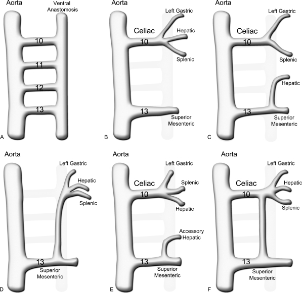
The arterial and venous circulation of the bowel is complex and is characterized by marked redundancy of multiple interconnecting branches, which provides a rich blood supply to aid in the digestive process and also serves to protect the bowel from potential ischemia or infarction. As a result of this circulatory pattern, anatomic variants and extensive collateral pathways are common. A thorough knowledge of both the arterial and venous mesenteric circulation, including normal, variant, and collateral anatomy, is necessary for the appropriate evaluation and management of the various disease processes that may affect the vascular supply of the gastrointestinal system.

Keywords: Mesenteric circulation; mesenteric collateral pathways; variant arterial anatomy.


Figures

Figure 1
Figure 1
Embryology of normal and variant mesenteric vascular anatomy. (A) In the primitive vasculature, the 10th to 13th vitelline arteries communicate between the aorta and a primitive ventral anastomotic artery. (B) Normally the ventral anastomosis and the 11th and 12th vitelline arteries regress, leaving the 10th root as the celiac trunk and the 13th as the superior mesenteric artery (SMA). (C) With replacement of the hepatic artery to the SMA, there is incomplete regression of the ventral anastomosis, forming a hepatomesenteric trunk. (D) A celiacomesenteric trunk occurs when the 10th to 12th vitelline arteries regress and a large portion of the ventral anastomosis persists to connect the celiac artery and branches to the SMA. (E) A partially replaced or accessory hepatic artery occurs in similar fashion to a completely replaced hepatic artery, through failure of a portion of the ventral anastomosis to regress. (F) The arc of Buhler results from persistence of the ventral anastomosis, connecting the celiac and SMA, despite regression of the 11th and 12th vitelline arteries.
Figure 2
Figure 2
Digital subtraction angiography arteriogram shows the common hepatic (medium white arrow), splenic (large white arrow) and left gastric (small black arrow) arteries as the three divisions of the celiac trunk. After the origin of the gastroduodenal artery (GDA; medium black arrow) the proper hepatic artery (large black arrow) courses and divides into the right (small white arrow) and left (white arrowhead) hepatic arteries. The GDA terminates as the right gastroepiploic artery (medium black arrowheads). Note that there are accessory left gastric artery branches (small black arrow-heads).
Figure 3
Figure 3
Selective arteriogram shows variant anatomy in which the entire middle colic artery (large black arrow) arises from the dorsal pancreatic artery (large black arrowhead) and then divides into the right (small white arrows) and left (small black arrows) branches to supply the entire transverse colon (small white arrowheads). The transverse pancreatic artery (small black arrowheads) also arises from the dorsal pancreatic artery and has multiple anastomoses with small branches (short black arrows) that arise from the splenic artery.
Figure 4
Figure 4
Selective common hepatic arteriogram shows the gastroduodenal (GDA) artery (small white arrow) and its terminal branch, the gastroepiploic artery (small white arrowheads). After yielding the GDA, the common hepatic becomes the proper hepatic artery (large black arrowhead), which then divides into the right (small black arrows) and left (small black arrowheads) hepatic arteries. The right gastric artery (large white arrows) arises from the proper hepatic artery, but may also arise from the left hepatic artery.
Figure 5
Figure 5
Selective arteriogram shows the left gastric artery (large white arrow) supplying the gastric cardia and retrograde filling of the right gastric artery (large white arrowheads) and opacification of a portion of the left hepatic artery (small white arrow) from which the right gastric artery arises. There is also retrograde filling of the gastroepiploic artery (small white arrowheads) and opacification of a short segment of the gastroduodenal artery (small black arrow), from which the right gastroepiploic artery originates.
Figure 6
Figure 6
Selective superior mesenteric artery (SMA; large white arrow) arteriogram shows the middle colic artery (large black arrowhead) divides into left (small black arrowheads) and right branches. Multiple jejunal and ileal branches (small white arrowheads) and the right colic artery (small white arrow) arise before the ileocolic artery (small black arrow), which is the terminal branch of the SMA.
Figure 7
Figure 7
Selective arteriogram shows the IMA (large white arrow) yields the left colic artery (small white arrows) which has an ascending division that anastomoses with the left branch of the middle colic artery (small black arrows) in the splenic flexure region. The terminal branch of the IMA is the superior rectal (hemorrhoidal) artery (white arrowhead). Note the marginal artery of Drummond (small black arrowheads) that courses along the mesenteric border of the colon.
Figure 8
Figure 8
Computed tomography angiogram shows the splenic vein (small white arrows) joining the superior mesenteric vein (large white arrow) to form the portal vein (white arrowhead), which bifurcates into the right (black arrowhead) and left (large black arrow) portal veins. Note that the right (small black arrowhead) and left (small black arrows) hepatic arteries course with the portal vein divisions into the liver to supply their respective anatomic territories.
Figure 9
Figure 9
Magnetic resonance angiogram shows a replaced right hepatic artery (large white arrow) arising from the superior mesenteric artery (large arrowhead). The left hepatic artery (small white arrow) arises normally from the celiac trunk, as does the left gastric artery (small arrowhead).
Figure 10
Figure 10
Angiogram of a combined celiacomesenteric trunk is seen in both frontal (A) and lateral (B) projections. The celiac vessel courses cephalad (white arrow) and the superior mesenteric artery courses caudally (white arrowhead). This anatomic variant occurs in <1% of individuals.
Figure 11
Figure 11
Selective inferior mesenteric artery (IMA) arteriogram shows the arc of Riolan (large white arrow) arising from the IMA (white arrowhead) and coursing medially in the mesentery to anastomose with the right (large black arrow) and left (small black arrow) branches of the middle colic artery. The marginal artery of Drummond (small white arrows) courses along the mesenteric border of the colon. It is somewhat inconstant and less developed in this patient.

References

    1. Reuter S R, Redman H C, Cho K J. Gastrointestinal Angiography. 3rd ed. Philadelphia, PA: WB Saunders; 1986.
    1. Kaufman J A, Lee M J. Vascular and Interventional Radiology: The Requisites. St. Louis, MO: Mosby; 2004.
    1. Petscavage J M, Maldjian P. Celiomesenteric trunk: two variants of a rare anomaly. Australas Radiol. 2007;51(Suppl):B306–B309. - PubMed
    1. Iezzi R, Cotroneo A R, Giancristofaro D, Santoro M, Storto M L. Multidetector-row CT angiographic imaging of the celiac trunk: anatomy and normal variants. Surg Radiol Anat. 2008;30(4):303–310. - PubMed
    1. McNulty J G, Hickey N, Khosa F, O'Brien P, O'Callaghan J P. Surgical and radiological significance of variants of Bühler's anastomotic artery: a report of three cases. Surg Radiol Anat. 2001;23(4):277–280. - PubMed