

Segmental arterial mediolysis
- PMID: 21326567
- PMCID: PMC3036489
- DOI: 10.1055/s-0029-1225666
Segmental arterial mediolysis
Abstract
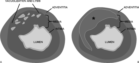
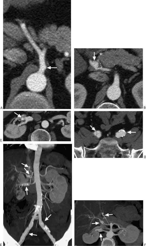
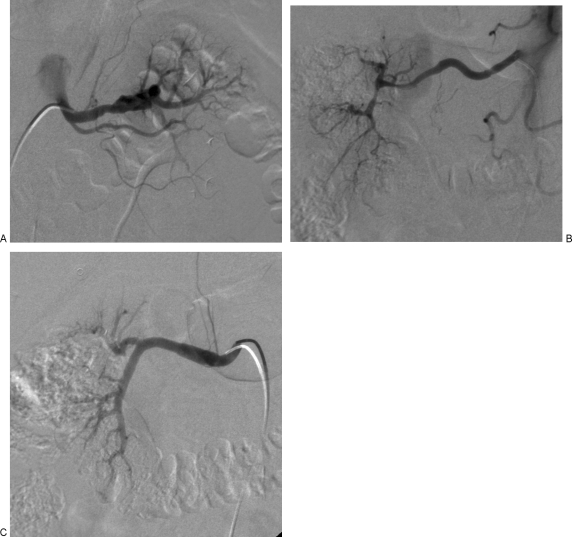
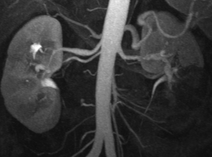
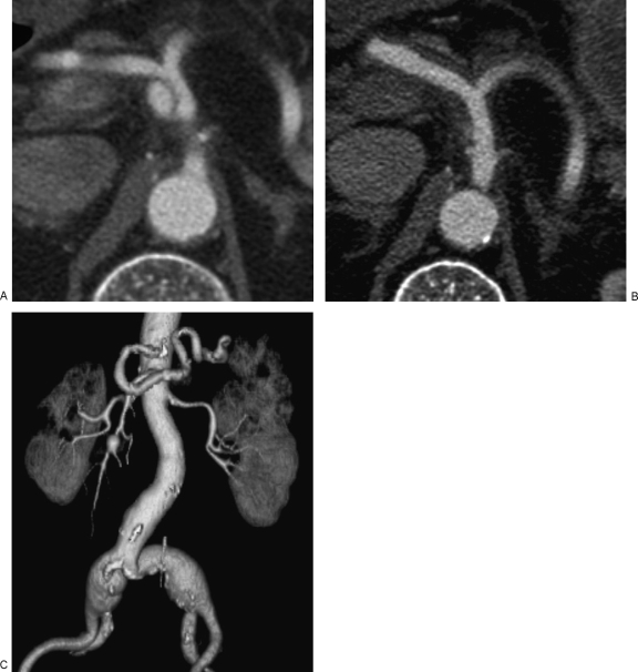
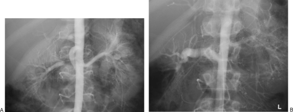
Segmental arterial mediolysis (SAM) is a nonatherosclerotic, noninflammatory arteriopathy, which is characterized by dissecting aneurysms resulting from lysis of the outer media of the arterial wall. The most common presentation is abdominal pain and hemorrhage in the elderly. Computed tomography (CT) and angiography imaging findings overlap with various vasculitides and include segmental changes of aneurysm and stenosis. A key distinguishing feature is the presence of dissections, the principle morphologic expression of SAM. Differentiation and exclusion of an inflammatory arteritis is crucial in appropriate management, as immunosuppressants generally used for treatment of vasculitis may be ineffective or even worsen the vasculopathy. Although the disease can be self-limiting without treatment or with conservative medical therapy, the acute process carries a 50% mortality rate and may necessitate urgent surgical and/or endovascular therapy. Prompt recognition and diagnosis are therefore of utmost importance in appropriate management of this rare entity.
Keywords: Segmental arterial mediolysis; imaging findings.
Figures
Similar articles
-
Segmental arterial mediolysis.Cardiovasc Intervent Radiol. 2014 Jun;37(3):604-12. doi: 10.1007/s00270-014-0859-4. Epub 2014 Feb 20. Cardiovasc Intervent Radiol. 2014. PMID: 24554198 Review.
-
Segmental arterial mediolysis: a case of mistaken hemorrhagic pancreatitis and review of the literature.JOP. 2014 Jan 10;15(1):72-7. doi: 10.6092/1590-8577/2036. JOP. 2014. PMID: 24413790 Review.
-
Segmental Arterial Mediolysis: An Unusual Case Mistaken to be a Strangulated Hernia.WMJ. 2017 Aug;116(3):173-176. WMJ. 2017. PMID: 29323836 Free PMC article.
-
Segmental Arterial Mediolysis: A Case Study and Review of the Literature in Accurate Diagnosis and Management.Vasc Specialist Int. 2019 Sep;35(3):174-179. doi: 10.5758/vsi.2019.35.3.174. Epub 2019 Sep 30. Vasc Specialist Int. 2019. PMID: 31620405 Free PMC article.
-
Segmental Arterial Mediolysis and its Mimickers: A Case Report and Review of the Literature.Eur J Case Rep Intern Med. 2023 Oct 16;10(11):004085. doi: 10.12890/2023_004085. eCollection 2023. Eur J Case Rep Intern Med. 2023. PMID: 37920230 Free PMC article.
Cited by
-
Clinically Suspected Segmental Arterial Mediolysis of the Splanchnic Arteries: A Report of 2 Rare Cases.Am J Case Rep. 2021 Apr 8;22:e929013. doi: 10.12659/AJCR.929013. Am J Case Rep. 2021. PMID: 33830972 Free PMC article.
-
Potentially stress-induced acute splanchnic segmental arterial mediolysis with a favorable spontaneous outcome.J Vasc Surg Cases Innov Tech. 2017 Mar 6;3(1):26-29. doi: 10.1016/j.jvscit.2016.09.002. eCollection 2017 Mar. J Vasc Surg Cases Innov Tech. 2017. PMID: 29349369 Free PMC article.
-
Spontaneous isolated dissection of the superior mesenteric artery and aneurysm formation resulting from segmental arterial mediolysis: a case report.Diagn Pathol. 2017 Oct 16;12(1):74. doi: 10.1186/s13000-017-0664-x. Diagn Pathol. 2017. PMID: 29037200 Free PMC article.
-
Endovascular treatment of gastroduodenal artery pseudoaneurysm in a low-risk young female: Case report with literature review.Int J Surg Case Rep. 2025 Jun;131:111346. doi: 10.1016/j.ijscr.2025.111346. Epub 2025 Apr 23. Int J Surg Case Rep. 2025. PMID: 40286689 Free PMC article.
-
Unexpected Angiography Findings and Effects on Management.J Clin Imaging Sci. 2016 Sep 1;6:33. doi: 10.4103/2156-7514.189727. eCollection 2016. J Clin Imaging Sci. 2016. PMID: 27688932 Free PMC article.
References
-
- Slavin R E, Gonzalez-Vitale J C. Segmental mediolytic arteritis: a clinical pathologic study. Lab Invest. 1976;35:23–29. - PubMed
-
- Gruenwald P. Necrosis in the coronary arteries of newborn infants. Am Heart J. 1949;38(6):889–897. - PubMed
-
- Slavin R E, Cafferty L, Cartwright J., Jr Segmental mediolytic arteritis. A clinicopathologic and ultrastructural study of two cases. Am J Surg Pathol. 1989;13(7):558–568. - PubMed
-
- Slavin R E, Saeki K, Bhagavan B, Maas A E. Segmental arterial mediolysis: a precursor to fibromuscular dysplasia? Mod Pathol. 1995;8(3):287–294. - PubMed
-
- Armas O A, Donovan D C. Segmental mediolytic arteritis involving hepatic arteries. Arch Pathol Lab Med. 1992;116(5):531–534. - PubMed
