

Pelvic congestion syndrome: diagnosis and treatment
- PMID: 21326577
- PMCID: PMC3036528
- DOI: 10.1055/s-0028-1102998
Pelvic congestion syndrome: diagnosis and treatment
Abstract
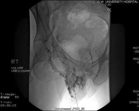
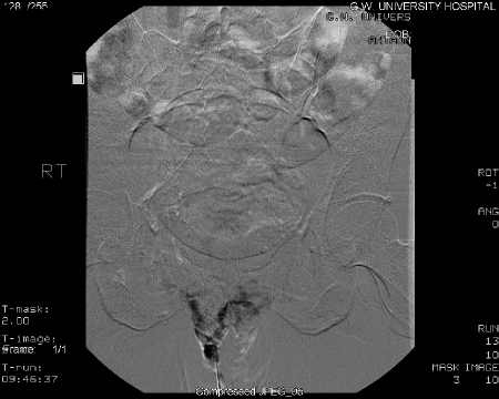
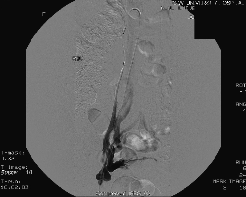
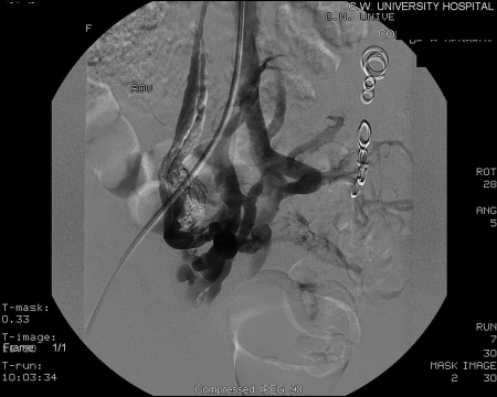
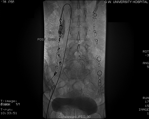
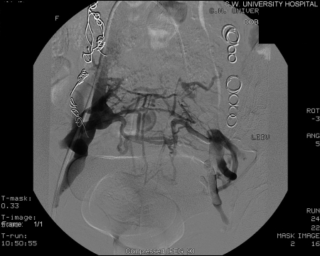
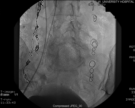
The diagnosis of pelvic congestion syndrome (PCS) continues to challenge all physicians involved especially those in such specialties as anesthesia, gastroenterology, general surgery, obstetrics and gynecology, and interventional radiology. When other pelvic pathology is ruled out, an interventional radiologist may be consulted for additional evaluation and treatment of PCS. A heightened awareness and clinical suspicion for the specific symptomatology and associated findings may bring about a more rapid progression toward treatment. For most interventional radiologists who treat PCS patients, magnetic resonance imaging/MR venography (MRI/MRV), diagnostic venogram, and embolotherapy are at the center of diagnosis and treatment of PCS.
Keywords: Chronic pelvic pain; congestion syndrome; embolization; ovarian vein embolotherapy; pelvic; pelvic varices.
Figures


References
-
- Robinson J C. Chronic pelvic pain. Curr Opin Obstet Gynecol. 1993;5:740–743. - PubMed
-
- Taylor H C. Vascular congestion and hyperemia: their effects on structure and function in the female reproductive system. Am J Obstet Gynecol. 1949;57:637–653. - PubMed
-
- Stones R W. Pelvic vascular congestion: half a century later. Clin Obstet Gynecol. 2003;46(4):831–836. - PubMed
-
- Park S J, Lim J W, Ko Y T, et al. Diagnosis of pelvic congestion syndrome using transabdominal and transvaginal sonography. AJR Am J Roentgenol. 2004;182(3):683–688. - PubMed
-
- Beard R W, Highman J H, Pearce S, et al. Diagnosis of pelvic varicosities in women with chronic pelvic pain. Lancet. 1984;2:946–949. - PubMed
LinkOut - more resources
Full Text Sources
Other Literature Sources
Medical
