. 2011 Feb 4;6(2):e14658.
doi: 10.1371/journal.pone.0014658.

Two-component system RgfA/C activates the fbsB gene encoding major fibrinogen-binding protein in highly virulent CC17 clone group B Streptococcus

Affiliations

Two-component system RgfA/C activates the fbsB gene encoding major fibrinogen-binding protein in highly virulent CC17 clone group B Streptococcus

Rim Al Safadi et al. PLoS One. .

Abstract

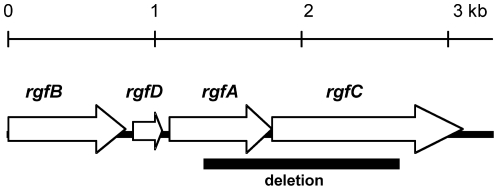
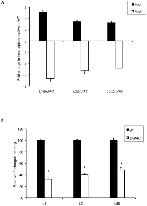
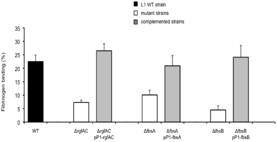
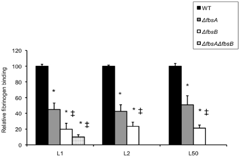
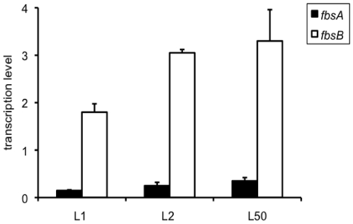
Group B streptococcus (GBS) strains with the highest ability to bind to human fibrinogen belong to the highly invasive clonal complex (CC) 17. To investigate the fibrinogen-binding mechanisms of CC17 strains, we determined the prevalence of fibrinogen-binding genes (fbsA and fbsB), and fbs regulator genes (rogB encoding an fbsA activator, rovS encoding an fbsA repressor and rgf encoding a two-component system [TCS] whose role on fbs genes was not determined yet) in a collection of 134 strains representing the major CCs of the species. We showed that specific gene combinations were related to particular CCs; only CC17 strains contained the fbsA, fbsB, and rgf genes combination. Non polar rgfAC deletion mutants of three CC17 serotype III strains were constructed. They showed a 3.2- to 5.1-fold increase of fbsA transcripts, a 4.8- to 6.7-fold decrease of fbsB transcripts, and a 52% to 68% decreased fibrinogen-binding ability, demonstrating that the RgfA/RgfC TCS inhibits the fbsA gene and activates the fbsB gene. The relative contribution of the two fbs genes in fibrinogen-binding ability was determined by constructing isogenic fbsA, fbsB, deletion mutants of the three CC17 strains. The ability to bind to fibrinogen was reduced by 49% to 57% in ΔfbsA mutants, and by 78% to 80% in ΔfbsB mutants, suggesting that FbsB protein plays a greater role in the fibrinogen-binding ability of CC17 strains. Moreover, the relative transcription level of fbsB gene was 9.2- to 12.7-fold higher than that of fbsA gene for the three wild type strains. Fibrinogen-binding ability could be restored by plasmid-mediated expression of rgfAC, fbsA, and fbsB genes in the corresponding deletion mutants. Thus, our results demonstrate that a specific combination of fbs genes and fbs regulator genes account for the high fibrinogen-binding ability of CC17 strains that may participate to their enhanced invasiveness for neonates as compared to strains of other CCs.


Conflict of interest statement

Competing Interests: The authors have declared that no competing interests exist.

Figures

Figure 1
Figure 1. Schematic representation of the rgf locus.
Open reading frames, direction of transcription and approximate gene sizes are indicated . The segment that was deleted in ΔrgfAC mutant is represented as a heavy line below the genes.
Figure 2
Figure 2. Properties of ΔrgfAC mutant strains.
(A) Fold change in transcription levels of fbsA (filled boxes) and fbsB (open boxes) genes in the isogenic ΔrgfAC mutants as compared to the wild type L1, L2, and L50 strains (WT). The amount of transcripts of each gene was normalized to the amount of gyrA transcripts and expressed relative to the level of transcription in corresponding WT strain. Each experiment was performed at least three times. Boxes are means and bars are standard deviation of the means. (B) Binding ability to immobilized human fibrinogen of the isogenic ΔrgfAC mutants (open boxes) and the WT strains (filled boxes). Flat bottomed 96-well polystyrene plates were coated with 21 nM human fibrinogen and 5×10⁶ to 5×10⁸ CFU per ml were added for 90 min at 37°C. Binding ability was calculated from the ratio between the number of bound bacteria and the number of bacteria present in the inoculum. The level of fibrinogen binding of WT strains is arbitrarily reported as 100 and the fibrinogen-binding levels of the isogenic mutants are relative values. Each experiment was performed at least three times. Boxes are means and bars are standard deviation of the means. * indicates that the binding values of the mutant strains were significantly lower than the values of the corresponding WT strains, at a P value of <0.001.
Figure 3
Figure 3. Binding ability to immobilized human fibrinogen of the wild type (WT) L1 strain, and of isogenic mutant and complemented strains for rgfAC, fbsA, and fbsB genes.
Flat bottomed 96-well polystyrene plates were coated with 21 nM human fibrinogen and 5×10⁶ to 5×10⁸ CFU per ml were added for 90 min at 37°C. Binding ability was calculated from the ratio between the number of bound bacteria and the number of bacteria present in the inoculum. Each experiment was performed at least three times. Boxes are means and bars are standard deviation of the means. The binding values of the mutant strains were significantly lower, at a P value of <0.001, than the values of the L1WT strain and of the corresponding complemented strains carrying rgfAC, fbsA, and fbsB genes on the pP1 plasmid.
Figure 4
Figure 4. Binding ability to immobilized human fibrinogen of the wild type (WT) S. agalactiae strains and isogenic ΔfbsA, ΔfbsB, ΔfbsAΔfbsB deletion mutants.
Flat bottomed 96-well polystyrene plates were coated with 21 nM human fibrinogen and 5×10⁶ to 5×10⁸ CFU per ml were added for 90 min at 37°C. Binding ability was calculated from the ratio between the number of bound bacteria and the number of bacteria present in the inoculum. The fibrinogen-binding level of WT L1, L2, and L50 strains is arbitrarily reported as 100 and the fibrinogen-binding levels of the various isogenic mutants are relative values. Each experiment was performed at least three times. Boxes are means and bars are standard deviation of the means. * indicates that the binding values of the mutant strains were significantly lower than the values of the corresponding WT strains, at a P value of <0.001. ‡ indicates that the binding values of the ΔfbsB and ΔfbsAΔfbsB mutant strains were significantly lower than the values of ΔfbsA mutant strains, at a P value of <0.001.
Figure 5
Figure 5. Transcription levels of fbsA and fbsB genes in three wild type CC17 strains.
The amount of transcripts of fbsA gene (filled boxes) and fbsB gene (open boxes) in L1, L2, and L50 wild type strains was normalized to the amount of gyrA transcripts. Each experiment was performed at least three times. Boxes are means and bars are standard deviation of the means.

References

    1. Farley MM. Group B streptococcal disease in nonpregnant adults. Clin Infect Dis. 2001;33:556–561. - PubMed
    1. Schuchat A. Epidemiology of group B streptococcal disease in the United States: shifting paradigms. Clin Microbiol Rev. 1998;11:497–513. - PMC - PubMed
    1. Bisharat N, Jones N, Marchaim D, Block C, Harding RM, et al. Population structure of group B streptococcus from a low-incidence region for invasive neonatal disease. Microbiology. 2005;151:1875–1881. - PubMed
    1. Chatellier S, Ramanantsoa C, Harriau P, Rolland K, Rosenau A, et al. Characterization of Streptococcus agalactiae strains by randomly amplified polymorphic DNA analysis. J Clin Microbiol. 1997;35:2573–2579. - PMC - PubMed
    1. Jones N, Bohnsack JF, Takahashi S, Oliver KA, Chan M, et al. Multilocus sequence typing system for group B streptococcus. J Clin Microbiol. 2003;41:2530–2536. - PMC - PubMed

Publication types

MeSH terms