

doi: 10.1055/s-2007-971180.
Lymphangiography: a case study
Affiliations
- PMID: 21326747
- PMCID: PMC3036359
- DOI: 10.1055/s-2007-971180
Item in Clipboard
Lymphangiography: a case study
Semin Intervent Radiol.
2007 Mar.
Abstract
Lymphatic leak is a rare but well-described complication of a multitude of surgeries, whose sequela may potentially be life threatening. For cases refractory to conservative management, surgical therapy has been the mainstay of treatment. Although radiology has always played a contributory role in the diagnosis of lymphatic leaks with lymphoscintigraphy and lymphangiography, minimally invasive management of lymphatic leaks by interventional radiologists has only been described in the last decade. We present a case of percutaneous disruption of the cisterna chyli to treat a lymphatic leak of the thoracic duct.
Keywords: Lymphangiogram; cisterna chili; lymphatic leak.
Figures
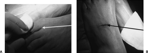
(A) Photograph of foot during subcutaneous injection of 75% methylene blue and 25% epinephrine into web space between digits 1 and 2 with 22-gauge needle (white arrow). (B) Photograph of feet following superficial incision along dorsum of foot to dissect out lymphatics (black arrow).
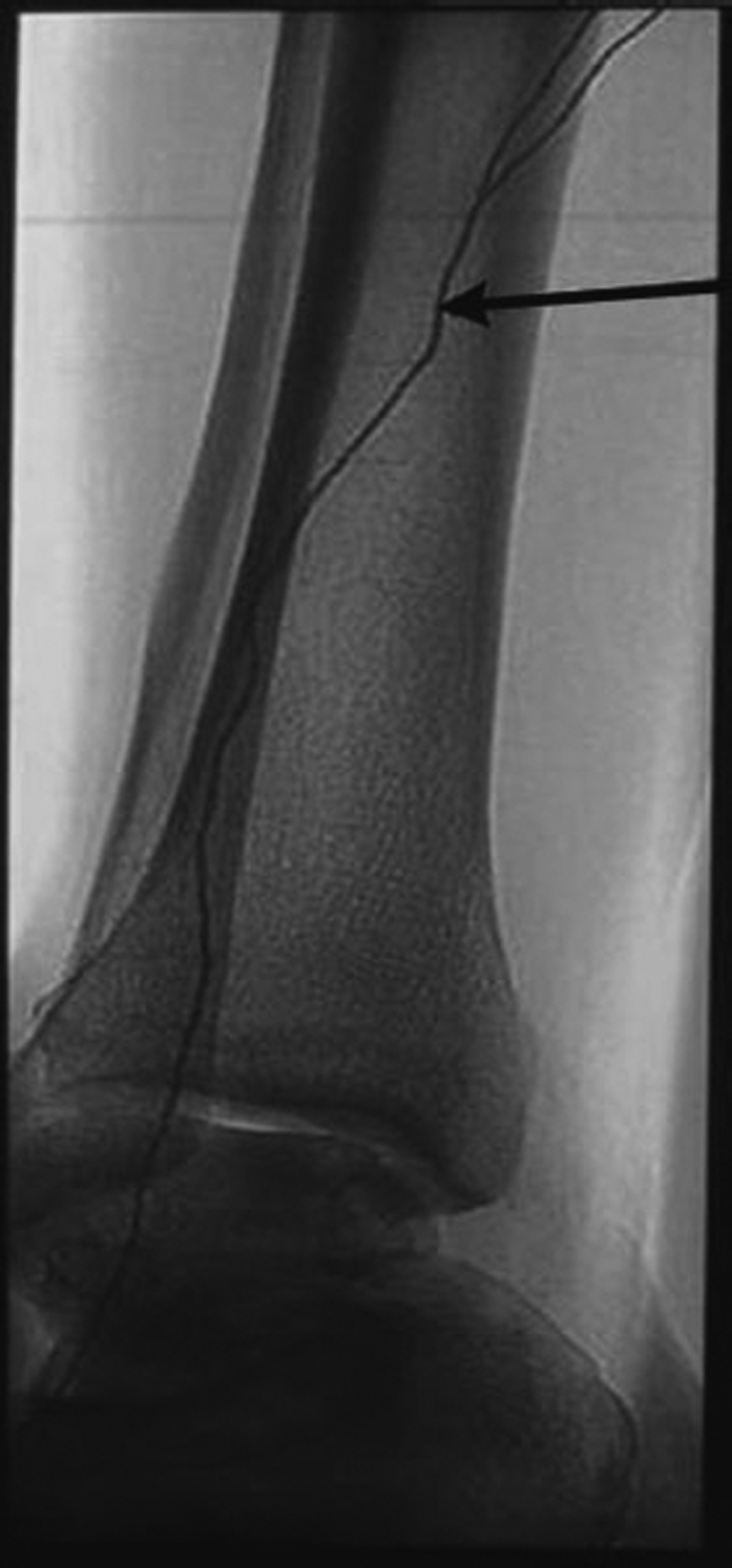
Lateral radiograph of ankle demonstrates Ethiodol tracking uniformly through the lymphatic system (arrow). In contrast, due to its oil base, Ethiodol forms globules in the vascular system, thereby differentiating the two.
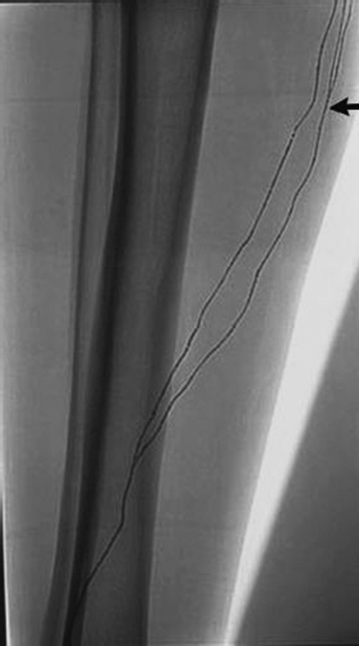
Intermittent spot radiograph of calf reveals opacified lymphatics (arrow) with cranial progression of Ethiodol.
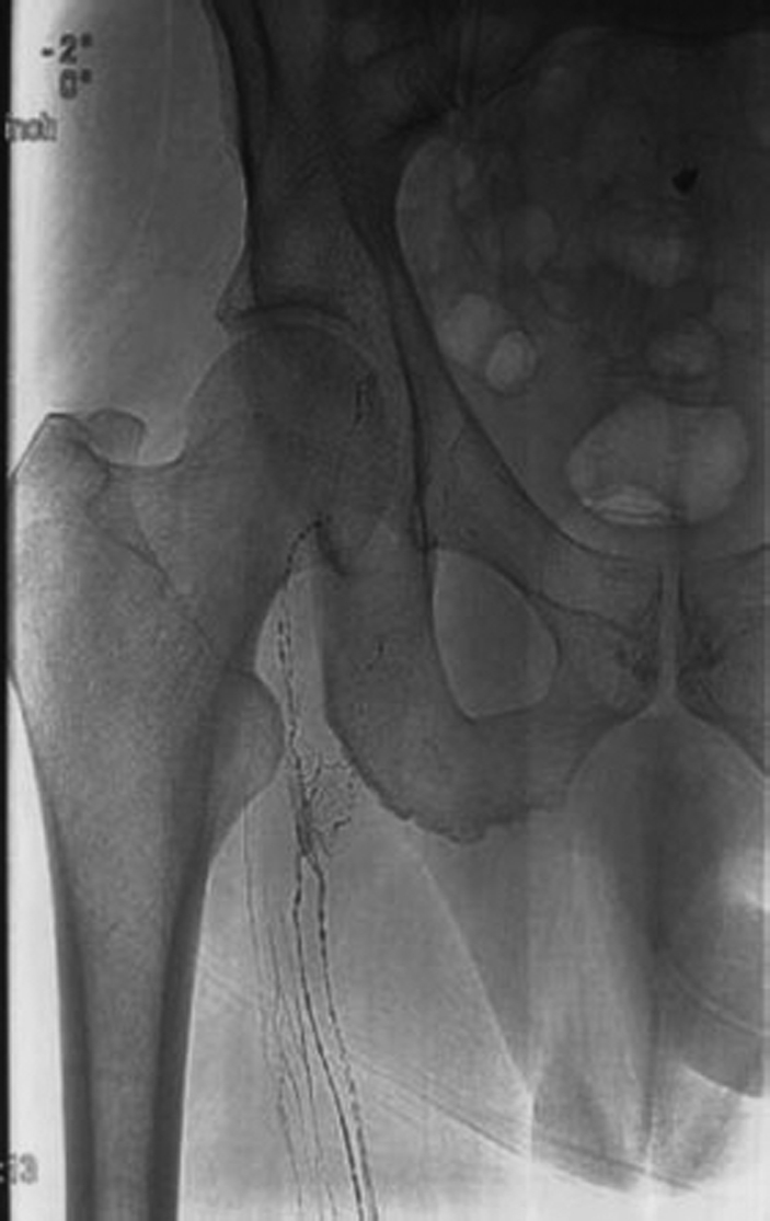
Radiograph of right upper thigh reveals cranial progression of Ethiodol.
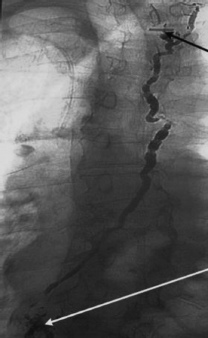
Chest radiograph reveals Ethiodol opacifying thoracic duct. Thoracic duct travels cranial and anterior to drain into subclavian vein. Black arrow points to surgical clip and site of leak. Ethiodol is left within the thoracic duct to promote sclerosis. Cisterna chyli has lost its normal globular appearance and is disrupted after ethanol ablation (white arrow).
References
-
- Barnacle A M, Kleidon T M. Lymphatic leak complicating central venous catheter insertion. Cardiovasc Intervent Radiol. 2005;28:839–840. - PubMed
-
- Porziella V, Cesario A, Margaritora S, Granone P. Role of pre-operative lymphangiogram and lympangioscintigraphy in the surgical management of spontaneous chylothorax. Eur J Cardiothorac Surg. 2006;30:813. - PubMed
-
- Merrigan B A, Winter D C, O'Sullivan G C. Chylothorax. Br J Surg. 1997;84:15–20. - PubMed
-
- Leibovitch I, Mor Y, Golomb J, Ramon J. The diagnosis and management of postoperative chylous ascites. J Urol. 2002;167:449–457. - PubMed
-
- Yamagami T, Masunami T, Kato T, et al. Spontaneous healing of chyle leakage after lymphangiography. Br J Radiol. 2005;78:854–857. - PubMed
