. 2006 Jun;23(2):150-5.
doi: 10.1055/s-2006-941445.

Complications of inferior vena caval filters

Affiliations

Complications of inferior vena caval filters

Thuong G Van Ha. Semin Intervent Radiol. 2006 Jun.

Abstract

Inferior vena caval filters have been shown to be effective in the prevention of pulmonary embolism, with low morbidity and mortality associated with their implantation. Awareness of potential complications can further decrease the risk of filter placement and lead to early detection and management of complications to improve clinical outcomes. The purpose of this article is to review the procedure-related and delayed complications associated with inferior vena caval filters.

Keywords: Inferior vena caval filters; complications; pulmonary embolism; thromboembolic disease.


Figures
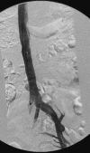
Figure 1
Figure 1
Proper IVC filter placement. The tip of the filter is at or near the level of the renal vein inflow.
Figure 2
Figure 2
A 39-year-old woman with hypercoagulable state presenting with DVT. (A) IVC-gram revealed extensive infrarenal IVC clot. (B) Fluoroscopic image showed that the filter was placed above the renal vein.
Figure 3
Figure 3
A 54-year-old man presented with DVT for IVC placement. (A) The first filter was deployed in a lumbar vein. (B) Another filter was placed in the infrarenal IVC.
Figure 4
Figure 4
A 48-year-old man with Recovery IVC filter (Bard, Covington, GA) placed for DVT prophylaxis. Approximately 1 year later, patient was admitted for unrelated reason. During this hospitalization, patient complained of palpitation and chest pain. (A) Chest X-ray revealed migration of the filter to the thorax. (B) Correlation with CT showed the filter in the right heart, straddling the tricuspid valve. Open-heart surgery was required for removal.
Figure 5
Figure 5
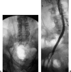
A 49-year-old man with bilateral lower extremity swelling presented for therapy. (A) Venogram showed extensive clot in the right iliac vein and thrombosed IVC with a Bird's Nest filter (Cook, Bloomington, IN) in position at the confluence of the iliac veins. (B) After thrombolysis and stenting, flow was restored through the iliac vein. The lower extremity edema resolved.
Figure 6
Figure 6
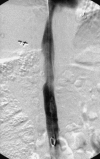
Venogram showed clots within the cone of the filter. Large clot burden within the filter cone is a contraindication to routine removal.
Figure 7
Figure 7
The Tulip filter (Cook, Bloomington, IN) is tilted and the hook is apposed to the IVC wall, preventing successful removal in this patient.

References

    1. Bick R L. Hereditary and acquired thrombophilia: preface. Semin Thromb Hemost. 1999;25:251–253. - PubMed
    1. Goldhaber S Z. Pulmonary embolism. N Engl J Med. 1998;339:93–104. - PubMed
    1. Athanasoulis C A, Kaufman J A, Halpern E F, Waltman A C, Geller S C, Fan C. Inferior vena caval filters: review of a 26-year single-center experience. Radiology. 2000;216:54–66. - PubMed
    1. Ferris E J, McCowan T C, Carver D K, McFarland D R. Percutaneous inferior vena caval filters: follow-up of seven designs in 320 patients. Radiology. 1993;188:851–856. - PubMed
    1. Crochet D P, Stora O, Ferry D, et al. Vena-Tech-LGM filter: long-term results of a prospective study. Radiology. 1993;188:857–860. - PubMed