. 2006 Sep;23(3):223-9.
doi: 10.1055/s-2006-948759.

Bronchial artery embolization for treatment of life-threatening hemoptysis

Affiliations

Bronchial artery embolization for treatment of life-threatening hemoptysis

January K Lopez et al. Semin Intervent Radiol. 2006 Sep.

Abstract

Massive hemoptysis is an emergent and life-threatening condition with a broad range of underlying causes. Fortunately, massive hemoptysis accounts for a minority of cases of hemoptysis, ~5%. Unlike hemorrhage in other areas of the body, the primary cause of death from pulmonary hemorrhage is most commonly asphyxiation rather than exsanguination. Given the limited capacity for the lung to preserve oxygen transfer in the setting of massive hemoptysis, a rapid and effective method for controlling hemorrhage is essential to minimize death in patients demonstrating respiratory compromise. Since its introduction in 1973, bronchial artery embolization has proven to be a safe and effective tool for the treatment of massive hemoptysis and is now considered the treatment of choice, with initial success rates ranging from 77 to 94%. The long-term control rate of hemoptysis ranges from 70 to 85% and is largely a function of the degree of inflammation and the natural progression of the underlying disease. This article reviews the current literature on bronchial artery embolization for the treatment of massive hemoptysis.

Keywords: Hemoptysis; bronchial artery embolization; embolization; massive hemoptysis.


Figures

Figure 1
Figure 1
Common trunk to bilateral bronchial arteries (arrow). A single bronchial artery is identified on each side.
Figure 2
Figure 2
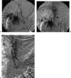
A patient with history of renal cell carcinoma, metastasizing to the mediastinum, presented with an erosive lesion into the left main stem bronchus. (A) Initial diagnostic angiogram demonstrated a right intercostal bronchial trunk (ICBT) with its origin at the inferior aspect of the distal aortic arch. This was noted to have direct feeding vessels to the subcarinal mass. A left bronchial artery (not shown) was interrogated and found to have no direct feeding supply to the mediastinal tumor. (B) The right ICBT was then embolized with a coaxial system using an outer 5F Cobra catheter (arrow) and an inner 3F microcatheter (arrowhead). Polyvinyl alcohol particles (500 to 700 μm) were utilized as the embolic agent. (C) Postembolization angiogram demonstrated cessation of flow (arrow) to the tumor mass as well as the right bronchial artery.
Figure 3
Figure 3
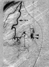
A 62-year-old woman with chronic obstructive pulmonary disease and bronchiectasis presented with recurrent hemoptysis, status post prior bronchial artery embolization. Thoracic aortogram revealed no bronchial artery. (A) The left internal mammary artery (ima) was evaluated and shown to supply tortuous, irregular vessels (arrowheads) to the left lingula. (B) These abnormal branches were embolized with polyvinyl alcohol particles using a coaxial system with an inner microcatheter (arrows).
Figure 4
Figure 4
A 22-year old woman with a history of lymphangiomas and hemangiomas presented with recurrent hemoptysis. She had previously undergone right lower lobe resection. Evaluation of the pulmonary and bronchial arteries was unremarkable. An arteriovenous fistula (long arrow) was found involving the distal portion of a right internal mammary arterial branch (ima). It was uncertain whether the fistula connected to the pulmonary artery or vein (arrowheads). This was likely postsurgical in nature.
Figure 5
Figure 5
Injection at the origin of the right superior intercostal artery demonstrated an anterior spinal artery arising from it, with the characteristic hairpin loop appearance (arrows).
Figure 6
Figure 6
A 48-year-old male with a history of metastatic plasmacytoma presented with hemoptysis. A right intercostals artery was selectively cannulated at the level of the carina, supplying multiple areas of abnormal blush, including a peripheral area (arrows) feeding the pleural thickening.

References

    1. In: Fraser RS, Muller NL, Colman N, Pare PD, editor. Diagnosis of Diseases of the Chest. Philadelphia: WB Saunders; 1999. The clinical history and physical examination. pp. 379–403.
    1. de Gregorio M A, Medrano J, Mainar A, et al. Endovascular treatment of massive hemoptysis by bronchial artery embolization: short-term and long-term follow-up over a 15-year period. Arch Bronconeumol. 2006;42:49–56. - PubMed
    1. Corr P D. Bronchial artery embolization for life-threatening hemoptysis using tris-acryl microspheres: short-term result. Cardiovasc Intervent Radiol. 2005;28:439–441. - PubMed
    1. Ustunsoz B, Bozlar U, Ors F, Gurkok S, Balkan A, Ilica T. Bronchial artery embolization: experience with 10 cases. Diagn Interv Radiol. 2006;12:43–46. - PubMed
    1. Yoon W, Kim J K, Kim Y H, Chung T W, Kang H K. Bronchial and nonbronchial systemic artery embolization for life-threatening hemoptysis: a comprehensive review. Radiographics. 2002;22:1395–1409. - PubMed