

Laparoscopy-assisted resection of tailgut cysts: report of a case
- PMID: 21326854
- PMCID: PMC3037990
- DOI: 10.1159/000322912
Laparoscopy-assisted resection of tailgut cysts: report of a case
Abstract
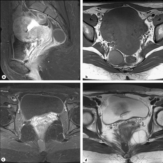
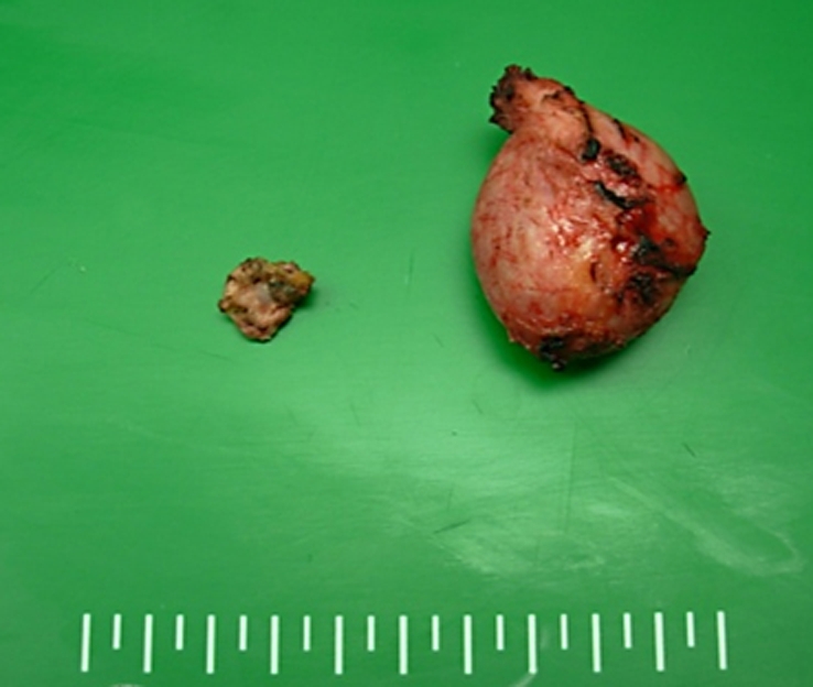
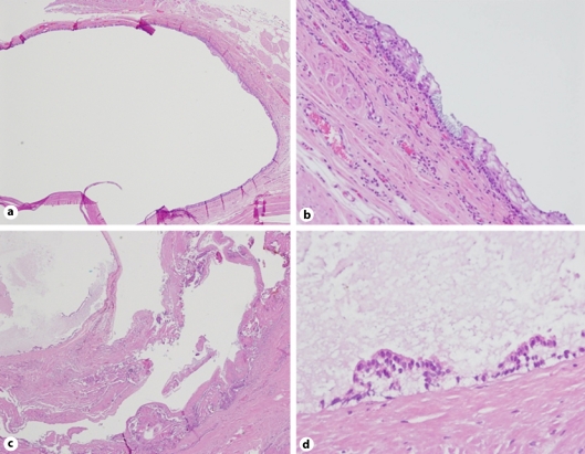
Tailgut cysts, or retrorectal cystic hamartomas, are rare congenital developmental lesions, most commonly located in the retrorectal space, and are more common in women. We present a case of retrorectal tailgut cyst managed using a laparoscopic approach. A 36-year-old woman presented with incidentally detected retrorectal tumors during evaluation for a gallbladder polyp. Her past medical history revealed that she had undergone cesarean section twice. The tumor marker CA 19-9 level was 42.52 U/ml. CT of the pelvis with contrast and pelvic MRI revealed a 3.9 × 3.3 cm well-defined, homogeneous cystic mass in the right presacral area, and a 2.5 × 1.5 cm cystic mass in the precoccygeal space. The patient underwent laparoscopic exploration with a preoperative diagnosis of tailgut cysts based on radiological findings. The operative time was 90 min including 30 min of subsequent laparoscopic cholecystectomy without placement of additional trocars. The surgical specimens consisted of two fragments of fibrofatty tissues, unilocular cystic masses. The final pathologic diagnosis was tailgut cysts with no evidence of malignancy. Postoperative recovery was uneventful, and the patient was discharged after 3 days. In conclusion, surgical resection is recommended in the management of retrorectal tailgut cyst to establish a definite diagnosis and to rule out malignancy. The laparoscopic approach is a feasible and safe option.
Keywords: Laparoscopic approach; Retrorectal mass; Surgical resection; Tailgut cyst.
Figures
References
-
- Rafindadi AH, Shehu SM, Ameh EA. Retrorectal cystic harmatoma (tailgut cyst) in an infant: case report. East Afr Med J. 1998;75:726–727. - PubMed
-
- Hjermstad BM, Helwig EB. Tailgut cysts. Report of 53 cases. Am J Clin Pathol. 1988;89:139–147. - PubMed
-
- Hutton KA, Benson EA. Case report: tailgut cyst – assessment with transrectal ultrasound. Clin Radiol. 1992;45:288–289. - PubMed
-
- Mourra N, Caplin S, Parc R, Flejou JF. Presacral neuroendocrine carcinoma developed in a tailgut cyst: report of a case. Dis Colon Rectum. 2003;46:411–413. - PubMed
-
- Schwarz RE, Lyda M, Lew M, Paz IB. A carcinoembryonic antigen-secreting adenocarcinoma arising within a retrorectal tailgut cyst: clinicopathological considerations. Am J Gastroenterol. 2000;95:1344–1347. - PubMed
Publication types
LinkOut - more resources
Full Text Sources
Miscellaneous
