. 2010 Jul;1(4):254-268.
doi: 10.4161/gmic.1.4.12778. Epub 2010 Jun 23.

Mucin-bacterial interactions in the human oral cavity and digestive tract

Affiliations

Mucin-bacterial interactions in the human oral cavity and digestive tract

Muriel Derrien et al. Gut Microbes. 2010 Jul.

Abstract

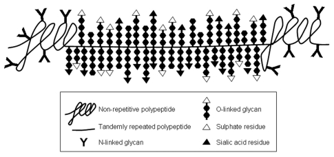
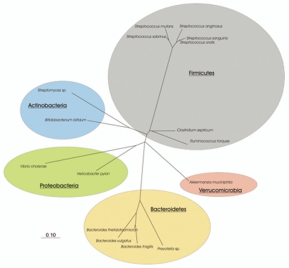
Mucins are a family of heavily glycosylated proteins that are the major organic components of the mucus layer, the protective layer covering the epithelial cells in many human and animal organs, including the entire gastro-intestinal tract. Microbes that can associate with mucins benefit from this interaction since they can get available nutrients, experience physico-chemical protection and adhere, resulting in increased residence time. Mucin-degrading microorganisms, which often are found in consortia, have not been extensively characterized as mucins are high molecular weight glycoproteins that are hard to study because of their size, complexity and heterogeneity. The purpose of this review is to discuss how advances in mucus and mucin research, and insight in the microbial ecology promoted our understanding of mucin degradation. Recent insight is presented in mucin structure and organization, the microorganisms known to use mucin as growth substrate, with a specific attention on Akkermansia muciniphila, and the molecular basis of microbial mucin degradation owing to availability of genome sequences.


Figures

Figure 1
Figure 1
Schematic representation of a mucin molecule. The part of the polypeptide where the O-linked glycans are concentrated is not drawn to scale. Based on electron micrographs of isolated mucin molecules, this glycopeptide comprises at least 70% of the length of the mucin molecule.
Figure 2
Figure 2
Schematic representation of the different associations of gel forming mucins with intestinal bacteria. Pathogens can be bound by proteolytically degraded mucin in the lumen (1), as mucins contain similar carbohydrate structures as found on the epithelium and thus be expelled. Similarly pathogens might be bound by the mucins in the mucus layer and therefore not reach the epithelium. When pathogens reach the epithelium they can bind to receptors (often carbohydrate structures) and might translocate across the epithelium (2). Normally, this will primarily occur when the mucus layer is gone as in severe pathology or concern microbes that can achieve mobility through the mucus layer, such as H. pylori in gastric mucus. Commensal bacteria can be present as planktonic bacteria in the lumen, live bound to the mucins in the mucus layer or live protected within the mucus without direct interactions (3). Commensal bacteria might compete with pathogens for binding sites on the mucins, thereby denying access to the pathogens (1 vs. 3). Some commensal as well as some pathogenic bacteria are able to proteolytically degrade mucins (4). Commensals, pathogens and probiotics are able to use mucins as energy source by degrading the O-glycans (5); the latter could happen within the mucus layer as well as in the lumen. The released monosaccharides are amongst others converted by commensal and probiotic bacteria into secondary metabolites (6), such as short chain fatty acids (SCFA), which are, e.g., essential for the colonic epithelium as an energy source. Please notice the scale of the components of the drawing. Mucin molecules (polymers) are several micrometers long, and are thus in the same range of size as the bacteria. In contrast, the epithelial cells are about 20 µm tall, whereas the mucus layer in, e.g., the colon can reach up to 800 µm in thickness.
Figure 3
Figure 3
Phylogenetic tree derived from the 16S rRNA sequence data of isolated and cultured mucin-degrading bacteria in the human digestive tract. Bar represents 10% divergence.

References

    1. Allen A. The structure and function of gastrointestinal mucus. In: Johnson LR, editor. Physiology of the Gastrointestinal Tract. Vol. 1. New York Raven Press; 1981. pp. 617–639.
    1. Johansson MEV, Phillipson M, Petersson J, Velcich A, Holm L, Hansson GC. The inner of the two Muc2 mucin-dependent mucus layers in colon is devoid of bacteria. PNAS. 2008;105:15064–15069. - PMC - PubMed
    1. Moore E. Physiology of intestinal and electrolyte absorption. American Gastroenterological Society. Baltimore: Milner-Fenwick; 1976.
    1. Allen A, Flemstrom G. Gastroduodenal mucus bicarbonate barrier: protection against acid and pepsin. Am J Physiol Cell Physiol. 2005;288:1–19. - PubMed
    1. Allen A. Mucus—a protective secretion of complexity. Trends Biochem Sci. 1983;8:169–173.

LinkOut - more resources