. 2011 Mar;135(3):305-15.
doi: 10.1007/s00418-011-0787-1. Epub 2011 Feb 16.

Myeloperoxidase and elastase are only expressed by neutrophils in normal and in inflamed liver

Affiliations

Myeloperoxidase and elastase are only expressed by neutrophils in normal and in inflamed liver

Ahmad Amanzada et al. Histochem Cell Biol. 2011 Mar.

Abstract

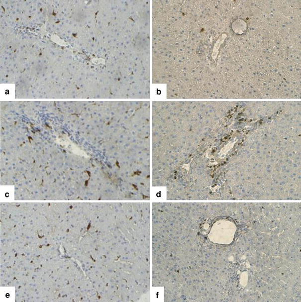
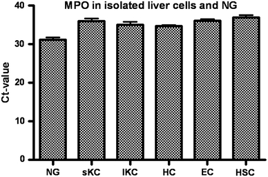
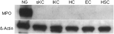
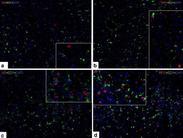
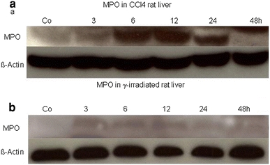
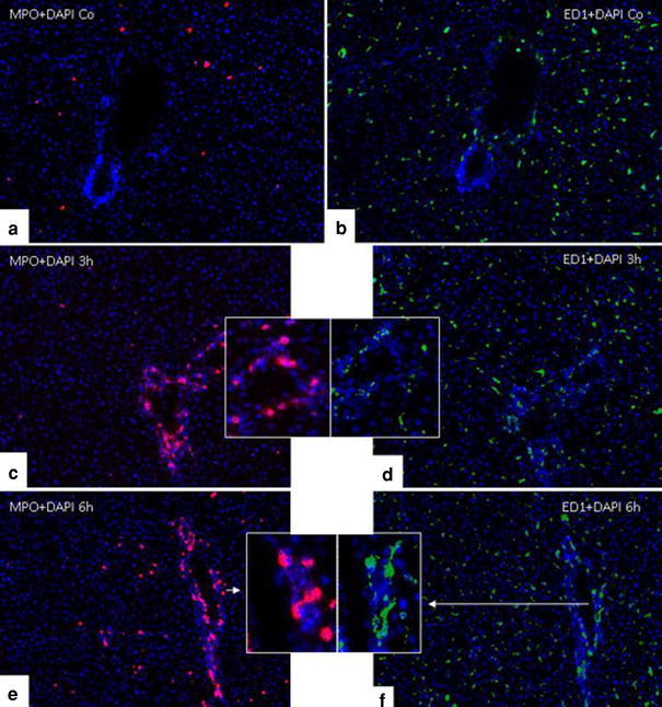
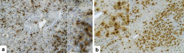
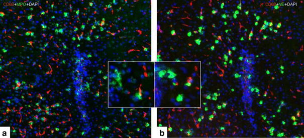
Myeloperoxidase (MPO) is involved in acute and chronic inflammatory diseases. The source of MPO in acute liver diseases is still a matter of debate. Therefore, we analysed MPO-gene expression on sections from normal and acutely damaged [carbon tetrachloride-(CCl(4)) or whole liver γ-Irradiation] rat liver by immunohistochemistry, real time PCR and Western blot analysis of total RNA and protein. Also total RNA and protein from isolated Kupffer cells, hepatic stellate cells, Hepatocytes, endothelial cells and neutrophil granulocytes (NG) was analysed by real time PCR and Western blot, respectively. Sections of acutely injured human liver were prepared for MPO and CD68 immunofluorescence double staining. In normal rat liver MPO was detected immunohistochemically and by immunofluorescence double staining only in single NG. No MPO was detected in isolated parenchymal and non-parenchymal cell populations of the normal rat liver. In acutely damaged rat liver mRNA of MPO increased 2.8-fold at 24 h after administration of CCl(4) and 3.3-fold at 3 h after γ-Irradiation and MPO was detected by immunofluorescence double staining only in elastase (NE) positive NGs but not in macrophages (ED1 or CD68 positive cells). Our results demonstrate that, increased expression of MPO in damaged rat and human liver is due to recruited elastase positive NGs.


Figures

Fig. 1
Fig. 1
Immunhistochemical detection of myeloperoxidase (MPO), neutrophil elastase (NE) and ED1 cells in rat liver 6 h after one single oral dose of CCl4-adminstration induced rat liver injury. MPO+ cells (a), ED1+ cells (b) and NE+ cells (c) in control rat liver. MPO+ cells (d), ED1+ cells (e) and NE+ cells (f) at 6 h after treatment. The lower panel shows MPO+ cells (g), ED1+ cells (h) and NE+ cells (i) at 24 h after treatment. Original magnification, ×100. G original magnification, ×200 173 × 116 mm (300 × 300 DPI)
Fig. 2
Fig. 2
MPO+ and ED1+ cells in rat liver after γ-Irradiation induced rat liver injury. ED1+ cells (a) and MPO+ cells (b) in control rat liver. ED1+ cells (c) and MPO+ cells (d) at 3 h after γ-Irradiation. ED1+ cells (e) and MPO+ cells (f) at 24 h after γ-Irradiation. Original magnification, ×100 173 × 174 mm (300 × 300 DPI)
Fig. 3
Fig. 3
MPO-gene expression in different cells analyzed by amplification of total RNA extracted from isolated cell populations of normal rat liver. Comparison of C t values of MPO in NG, small (sKC) and large KC (lKC), hepatocytes (HC), endothelial cells (EC) and hepatic stellate cells (HSC). Results were obtained by real time PCR analysis of total RNA. Results represent three experiments (in duplicate) and mean ± SEM values are shown for each cell type 46 × 34 mm (300 × 300 DPI)
Fig. 4
Fig. 4
Western blot analysis of total protein of NG, small KC’s (sKC), large KC’s (lKC), hepatocytes (HC), endothelial cells (EC) and hepatic stellate cells (HSC). Cells were isolated and cultured for 24 h. Protein was then extracted, 20 g of total protein were separated by SDS–PAGE, and blotted onto PVDF-membranes. The membranes were subsequently incubated with the antibodies against MPO (upper panel) and s-actin (lower panel). The molecular weight of MPO is 59 and 13.5 kDa. In addition, autocatalytic products (AP) can be seen at 40 and 20 kDa (data not shown). The molecular weight of β-actin is 42 kDa 129 × 41 mm (300 × 300 DPI)
Fig. 5
Fig. 5
Double staining of liver sections with monoclonal antibodies directed against MPO or NE (red) and monoclonal antibody against ED1 (green) followed by fluorescence immunodetection in sections of rat liver at different time points after CCl4-administration. a NE+ or ED1+ cells in CCl4-induced liver injury at 0 h. b MPO+ or ED1+ cells in CCl4-induced liver injury at 0 h. c NE+ or ED1+ cells in CCl4-induced liver injury 24 h after administration. d MPO+ or ED1+ cells in CCl4-induced liver injury 24 h after administration. Original magnification, ×100 173 × 130 mm (300 × 300 DPI)
Fig. 6
Fig. 6
Fold change of mRNA expression of MPO (a) and NE (b) after CCl4-administration induced rat liver injury at different time points. Real time PCR was normalized by using two housekeeping genes: β-actin and 18 s RNA. Results represent mean ± SEM value of three experiments (in duplicate) compared with controls for each time point 173 × 62 mm (600 × 600 DPI)
Fig. 7
Fig. 7
Western blot analysis of MPO of total protein in acute liver injury induced by CCl4 (a) and γ-Irradiation (b). Protein was extracted from livers of control (Co) rats or 3, 6, 12, 24 and 48 h after one single oral dose of CCl4-adminstration or γ-Irradiation induced rat liver injury. 20 μg of total protein were separated by SDS–PAGE, and blotted onto PVDF-membranes. The membranes were subsequently incubated with the antibodies against MPO (upper panel) and β-actin (lower panel). The molecular weight of the MPO is 59 and 13.5 kDa. In addition, autocatalytic products (AP) can be seen at 40 and 20 kDa (data not shown). The molecular weight of β-actin is 42 kDa 160 × 90 mm (300 × 300 DPI)
Fig. 8
Fig. 8
Immunofluorescence staining of liver sections after γ-Irradiation at different time points with monoclonal antibodies directed against MPO (red) and monoclonal antibody against ED1 (green). MPO+ cells (a) or ED1+ cells (b) in control liver. MPO+ cells (c) and ED1+ cells (d) at 3 h after γ-Irradiation. MPO+ cells (e) and ED1+ cells (f) at 6 h after γ-Irradiation. Original magnification, ×100 15 × 16 mm (600 × 600 DPI)
Fig. 9
Fig. 9
Fold change of mRNA expression of MPO (a) and NE (b) after γ-Irradiation induced rat liver injury at different time points. Real time PCR was normalized by using two housekeeping genes: β-actin and 18 s RNA. Results represent mean ± SEM value of three experiments (in duplicate) compared with controls for each time point 173 × 62 mm (600 × 600 DPI)
Fig. 10
Fig. 10
Fold change of mRNA expression of CD11b after CCl4-administration and γ-Irradiation induced rat liver injury at different time points. Real time PCR was normalized by using two housekeeping genes: β-actin and 18 s RNA. Results represent mean ± SEM value of three experiments (in duplicate) compared with controls for each time point 173 × 60 mm (600 × 600 DPI)
Fig. 11
Fig. 11
Indirect immunodetection of MPO (a) or NE (b) in sections of acute alcohol-toxic injured human liver. Sections were stained with antibodies against MPO or NE followed by peroxidase staining. Original magnification, ×100 173 × 49 mm (300 × 300 DPI)
Fig. 12
Fig. 12
Double staining of acute alcohol-toxic injured human liver sections with antibodies directed against MPO and NE (green) and antibody against CD68 (red) followed by fluorescence immunodetection. MPO+ or CD68+ cells (a) and NE+ or CD68+ cells (b). Original magnification, ×100 46 × 20 mm (600 × 600 DPI)

References

    1. Akiyama Y, Miller PJ, Thurman GB, Neubauer RH, Oliver C, Favilla T, Beman JA, Oldham RK, Stevenson HC. Characterization of human blood monocyte subset with low peroxidase activity. J Clin Invest. 1983;72:1093–1095. doi: 10.1172/JCI111034. - DOI - PMC - PubMed
    1. Armbrust T, Ramadori G. Functional characterization of two different Kupffer cell populations of normal rat liver. J Hepatol. 1996;25:518–528. doi: 10.1016/S0168-8278(96)80212-1. - DOI - PubMed
    1. Armbrust T, Schwögler S, Zöhrens G, Ramadori G. C1 esterase inhibitor gene expression in rat Kupffer cells, peritoneal macrophages and blood monocytes: modulation by interferon gamma. J Exp Med. 1993;178:373–380. doi: 10.1084/jem.178.2.373. - DOI - PMC - PubMed
    1. Armburst T, Ramadori G. Mononuclear phagocytes of acutely injured rat liver abundantly synthesize and secrete fibronectin in contrast to Kupffer cells of normal liver. Biochem Biophys Res Commun. 1995;213:752–758. doi: 10.1006/bbrc.1995.2194. - DOI - PubMed
    1. Arnhold J, Mueller S, Arnold K, Sonntag KJ. Mechanisms of inhibition of chemiluminescence in the oxidation of luminol by sodium hypochlorite. Biolum Chemilum. 1993;8:307–313. doi: 10.1002/bio.1170080604. - DOI - PubMed

MeSH terms