Review
. 2011 Apr;22(2):229-36.
doi: 10.1016/j.semcdb.2011.02.010. Epub 2011 Feb 15.

Purinergic mechanisms in gliovascular coupling

Affiliations
Review

Purinergic mechanisms in gliovascular coupling

Dale A Pelligrino et al. Semin Cell Dev Biol. 2011 Apr.

Abstract

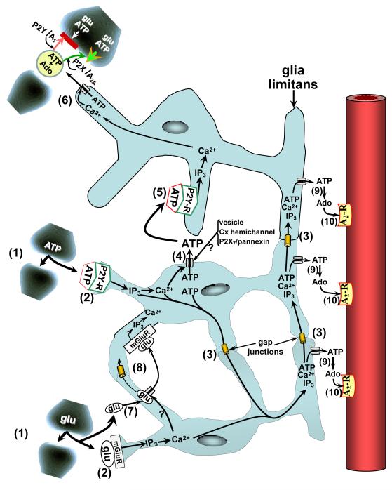
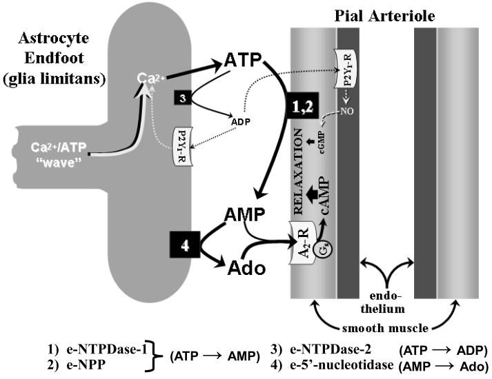
Regional elevations in cerebral blood flow (CBF) often occur in response to localized increases in cerebral neuronal activity. An ever expanding literature has linked this neurovascular coupling process to specific signaling pathways involving neuronal synapses, astrocytes and cerebral arteries and arterioles. Collectively, these structures are termed the "neurovascular unit" (NVU). Astrocytes are thought to be the cornerstone of the NVU. Thus, not only do astrocytes "detect" increased synaptic activity, they can transmit that information to proximal and remote astrocytic sites often through a Ca(2+)- and ATP-related signaling process. At the vascular end of the NVU, a Ca(2+)-dependent formation and release of vasodilators, or substances linked to vasodilation, can occur. The latter category includes ATP, which upon its appearance in the extracellular compartment, can be rapidly converted to the potent vasodilator, adenosine, via the action of ecto-nucleotidases. In the present review, we give consideration to experimental model-specific variations in purinergic influences on gliovascular signaling mechanisms, focusing on the cerebral cortex. In that discussion, we compare findings obtained using in vitro (rodent brain slice) models and multiple in vivo models (2-photon imaging; somatosensory stimulation-evoked cortical hyperemia; and sciatic nerve stimulation-evoked pial arteriolar dilation). Additional attention is given to the importance of upstream (remote) vasodilation; the key role played by extracellular ATP hydrolysis (via ecto-nucleotidases) in gliovascular coupling; and interactions among multiple signaling pathways.


Figures

FIGURE 1
FIGURE 1
Purine-linked coupling among components of the neurovascular unit (NVU). Neurovascular/gliovascular coupling begins with increased synaptic activity, represented by release of the neurotransmitters (gliotransmitters), ATP and glutamate (glu) (1). Engagement of metabotropic purinergic and glutamatergic receptors (P2Y-R and mGluR, respectively) on nearby astrocytes promotes PLC-mediated formation of IP3 and leads to Ca2+ release from intracellular stores (2). The Ca2+ and IP3 may be distributed among adjacent astrocytes and their processes either directly, via diffusion through intercellular gap junctional channels (3); or, indirectly, by Ca2+-dependent ATP release into the extracellular milieu via specialized release processes, including vesicles, connexin-linked hemichannels, and pannexin/P2X7-linked hemichannels (4). That ATP is then able to engage P2Y receptors on nearby astrocytes (5) resulting in elevations in cytosolic IP3 and Ca2+ levels in those cells. ATP also may move from astrocyte to astrocyte following the same gap junction diffusional routes as IP3 and Ca2+ (3). ATP efflux occurring in the vicinity of synapses (6) may lead to prevention (red bar) or enhancement (green arrowhead) of neurotransmitter release. This may occur via ATP interactions with P2Y or P2X receptors or, following ecto-nucleotidase-mediated conversion to adenosine (Ado), engagement of presynaptic A1 or A2A receptors [56]. Synaptic depression is thought to occur when P2Y and/or A1 receptors are involved; while facilitation may occur in association with P2X [57] and/or A2A [3] engagement. Following transporter-mediated uptake into astrocytes (7), glutamate may follow pathways similar to those of ATP (8). Efflux of ATP from the glia limitans should be accompanied by rapid ecto-nucleotidase-mediated formation of AMP, then Ado (9). Upon subsequent activation of pial arteriolar A2 receptors (10), vasodilation ensues (see fig. 2).
FIGURE 2
FIGURE 2
Postulated events thought to occur following the arrival of a Ca2+/ATP “wave” at the glia limitans and the subsequent Ca2+-dependent ATP release from the glia limitans. Preliminary findings indicated that, when ATP is topically applied to the cortical surface (mimicking SNS-associated increased ATP release from the glia limitans [see fig. 1]), the ensuing dilation of pial arterioles is mediated by products of ATP hydrolysis, the first step being a rapid ATP conversion to AMP, mediated through the actions of ecto-nucleoside triphosphate dihosphohydrolase-1 (e-NTPDase-1 (1) and ecto-pyrophosphatase/phosphodiesterase (e-NPP) (2), which are found on the surfaces of vascular cells and, perhaps, astrocytes as well [72,73]. Most of the AMP is converted to adenosine (Ado) via ecto-5′-nucleotidase (4), mainly found on astrocytes [73]. Pial arteriolar dilation arises from the cAMP generated following activation of vascular smooth muscle Gs-linked A2 receptors (A2-R). The principal A2-R ligands are Ado and (to a lesser degree) AMP. If extracellular ATP elevations are sufficiently high, modest generation of ADP may occur, allowing some ATP→ADP (i.e., ecto-ATPase) activity to be manifested [74], for example, via astrocytic e-NTPDase-2 (3). ADP is known to dilate pial arterioles through activation of purinergic P2Y1 receptors found both on the glia limitans and on vascular endothelium [75], with the latter representing a minor pathway linked to NO and cGMP generation.

References

    1. Halassa MM, Fellin T, Haydon PG. Tripartite synapses: roles for astrocytic purines in the control of synaptic physiology and behavior. Neuropharmacology. 2009;57:343–6. - PMC - PubMed
    1. Iadecola C, Nedergaard M. Glial regulation of the cerebral microvasculature. Nat Neurosci. 2007;10:1369–76. - PubMed
    1. Pelligrino DA, Xu HL, Vetri F. Caffeine and the Control of Cerebral Hemodynamics. J Alzheimers Dis. 2010;20(Suppl 1):S51–S62. - PMC - PubMed
    1. Koehler RC, Roman RJ, Harder DR. Astrocytes and the regulation of cerebral blood flow. Trends Neurosci. 2009;32:160–9. - PubMed
    1. Haydon PG, Carmignoto G. Astrocyte control of synaptic transmission and neurovascular coupling. Physiol Rev. 2006;86:1009–31. - PubMed

Publication types

Substances

LinkOut - more resources