. 2011 Apr 8;286(14):12066-74.
doi: 10.1074/jbc.M110.186882. Epub 2011 Feb 17.

Phosphorylation by CK2 enhances the rapid light-induced degradation of phytochrome interacting factor 1 in Arabidopsis

Affiliations

Phosphorylation by CK2 enhances the rapid light-induced degradation of phytochrome interacting factor 1 in Arabidopsis

Qingyun Bu et al. J Biol Chem. .

Abstract

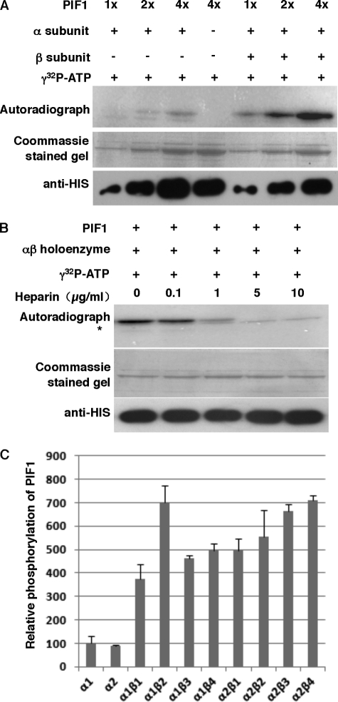
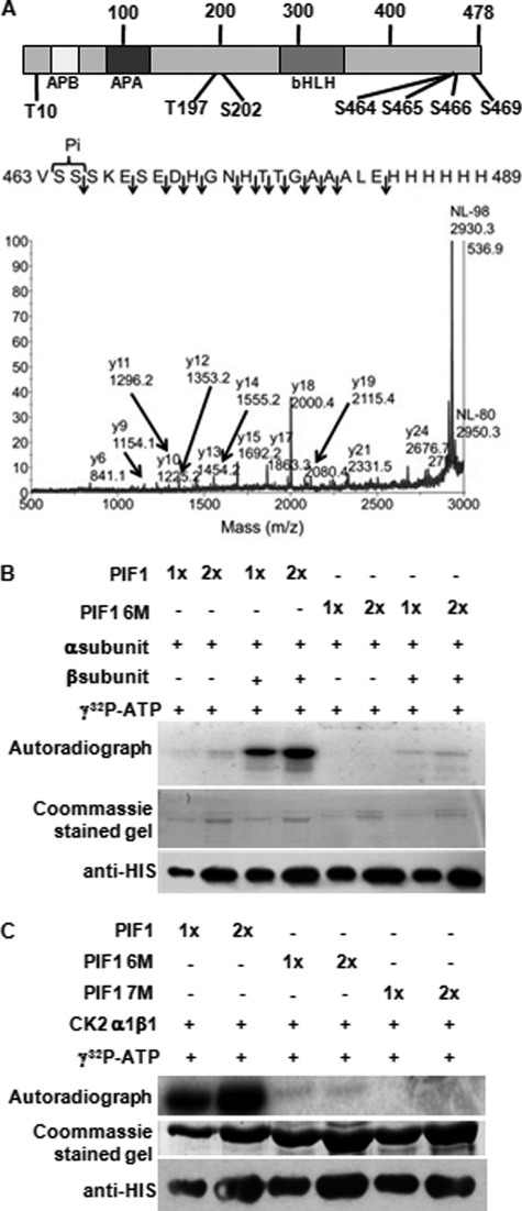
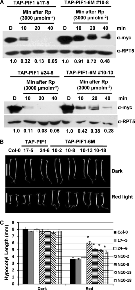
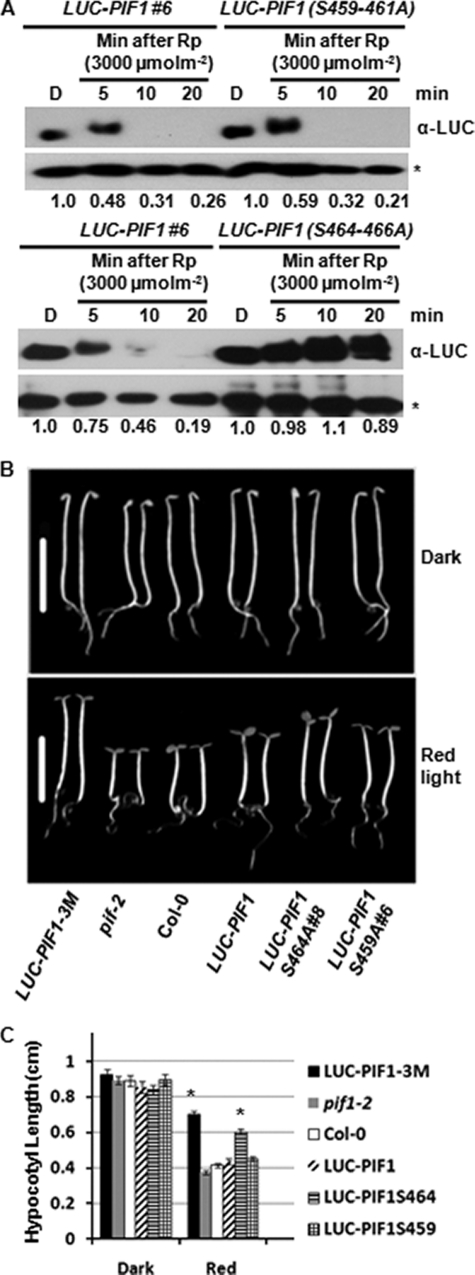
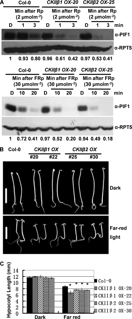
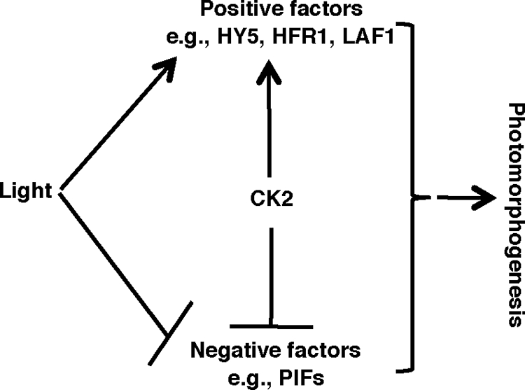
The phytochrome family of sensory photoreceptors interacts with phytochrome interacting factors (PIFs), repressors of photomorphogenesis, in response to environmental light signals and induces rapid phosphorylation and degradation of PIFs to promote photomorphogenesis. However, the kinase that phosphorylates PIFs is still unknown. Here we show that CK2 directly phosphorylates PIF1 at multiple sites. α1 and α2 subunits individually phosphorylated PIF1 weakly in vitro. However, each of four β subunits strongly stimulated phosphorylation of PIF1 by α1 or α2. Mapping of the phosphorylation sites identified seven Ser/Thr residues scattered throughout PIF1. Ser/Thr to Ala scanning mutations at all seven sites eliminated CK2-mediated phosphorylation of PIF1 in vitro. Moreover, the rate of degradation of the Ser/Thr to Ala mutant PIF1 was significantly reduced compared with wild-type PIF1 in transgenic plants. In addition, hypocotyl lengths of the mutant PIF1 transgenic plants were much longer than the wild-type PIF1 transgenic plants under light, suggesting that the mutant PIF1 is suppressing photomorphogenesis. Taken together, these data suggest that CK2-mediated phosphorylation enhances the light-induced degradation of PIF1 to promote photomorphogenesis.


Figures

FIGURE 1.
FIGURE 1.
CK2 phosphorylates PIF1 in vitro. A, phosphorylation of PIF1 by CK2 is enhanced by the β subunit. The autoradiogram shows that HIS-PIF1 was strongly phosphorylated by a recombinant CK2 holoenzyme in vitro. CK2 phosphorylation assays were performed in 20 μl of kinase assay mixtures that contained 50 mm Hepes-KOH (pH 7.6), 5 mm MgCl2, 2.4 mm DTT, 100 mm KCl, 0.2 mm γ-[32P]ATP (∼250 cpm/pmol), ∼1 pmol CK2 α or αβ holoenzyme, and ∼10–20 pmol PIF1. The reaction was incubated at 30 °C for 30 min and terminated by the addition of 4× SDS loading buffer. Samples were boiled for 3 min and separated on 10% SDS-PAGE gels. The gels were dried and exposed to a phosphorImager. B, autoradiogram showing that heparin effectively inhibited HIS-PIF1 phosphorylation by CK2 in a dosage-dependent manner. The kinase assays were performed as described in A. The asterisk indicates a nonspecific band. C, different subunit combinations of CK2 differentially phosphorylate PIF1 in vitro. The kinase assays were performed as described in A. Statistical analyses for significant differences are shown in supplemental Table S3.
FIGURE 2.
FIGURE 2.
PIF1 is phosphorylated by CK2 at multiple sites. A, diagram showing the CK2 phosphorylation sites identified in PIF1 (top panel). Identification of phosphorylation sites in PIF1 by MALDI mass spectrometry (bottom panel). MS/MS from the phosphopeptide 463 VSSSKESEDHGNHTTGAAALEHHHHHH 489 is shown. The spectrum is dominated by the neutral loss of 98-Da phosphoric acid that is characteristic of phosphopeptides. Fragments are observed from the C-terminal charge retention y ions up to y24, all without phosphorylation, indicating that the phosphorylation site is localized at Ser-464 and/or Ser-465. B, autoradiogram showing phosphorylation of wild-type HIS-PIF1 and HIS-PIF1-6M mutant proteins by purified recombinant CK2. In vitro phosphorylation of mutant PIF1 by CK2 is severely reduced. C, autoradiogram showing phosphorylation of wild-type HIS-PIF1, HIS-PIF1- 6M, and HIS-PIF1-7M mutant proteins by the recombinant CK2 α1β1 holoenzyme. In vitro phosphorylation of HIS-PIF1-7M by CK2 is completely eliminated.
FIGURE 3.
FIGURE 3.
CK2-mediated phosphorylation of PIF1 is necessary for rapid light-induced degradation of PIF1 in vivo. A, light-induced phosphorylation and degradation of a PIF1 containing six CK2 phosphorylation sites mutated (TAP-PIF1-6M) compared with wild-type TAP-PIF1. Two independent transgenic lines were compared with the corresponding wild-type TAP-PIF1 controls (top and bottom panels). α-RPT5 blots are shown as loading control. Numbers under the protein gel blots show the relative PIF1 level in the wild-type TAP-PIF1 and TAP-PIF1-6M transgenic lines. The PIF1 level in each dark samples is set as 1. B, TAP-PIF1-6M promotes hypocotyl growth under red light. Photographs of seedlings of various genotypes grown in the dark or under R light (7 μmol m−2s−1) for 4 days. Scale bars = 5 mm. Protein levels for various independent transgenic lines are shown (supplemental Fig. S2). C, bar graph showing the mean hypocotyl lengths for various genotypes as indicated. Seedlings were grown as described in B. Error bars represent mean ± S.E. (n ≥ 30). *, p < 0.05.
FIGURE 4.
FIGURE 4.
Ser-464–466 are necessary for the rapid light-induced degradation of PIF1. A, light-induced phosphorylation and degradation of a PIF1 containing either Ser-459–461 to Ala (top panel) or Ser-464–466 to Ala (bottom panel) compared with wild-type LUC-PIF1. The rate of light-induced degradation of LUC-PIF1(S464–466A) is strongly reduced compared with wild-type LUC-PIF1 (bottom panel). The asterisk indicates a cross-reacting band. The numbers under the protein gel blots show the relative PIF1 level in the wild-type LUC-PIF1, LUC-PIF1(S459–461A), and LUC-PIF1(S464–466A) transgenic lines. The PIF1 level in each dark samples is set as 1. B, LUC-PIF1(S464–466A) promotes hypocotyl growth under red light. Photographs of seedlings of various genotypes grown in the dark or under R light (7 μmol m−2s−1) for 4 days. Scale bars = 5 mm. A second allele of LUC-PIF1(S464–466A)#33 displaying a similar long hypocotyl phenotype under red light is shown in supplemental Fig. S4. C, bar graph showing the mean hypocotyl lengths of various genotypes as indicated. Seedlings were grown as described in B. Error bars represent mean ± S.E. (n ≥ 30). *, p < 0.05.
FIGURE 5.
FIGURE 5.
Degradation of PIF1 is faster in CK2 β1 and CK2 β2 overexpression lines under red light and far-red light. A, protein gel blots showing native PIF1 levels in wild-type and two independent CK2 β subunit overexpression lines. Four-day-old dark-grown seedlings were exposed to red pulse (Rp) (top panel) or far-red pulse (FRp) (bottom panel) at the indicated fluences and then incubated in the dark for the times indicated before harvesting for protein extraction. The numbers under the protein gel blots show the relative PIF1 level in the wild-type and CK2 β subunit overexpression lines. The PIF1 level in the wild-type Col-O dark sample is set as 1. α-RPT5 blots are shown as loading control. B, CK2 β1 and CK2 β2 overexpression lines are hypersensitive to FR light compared with the wild-type control. Photographs of seedlings of various genotypes grown in the dark or under FR light (0.4 μmol m−2s−1) for 4 days are shown. Scale bars = 5 mm. C, bar graph showing the mean hypocotyl lengths of various genotypes as indicated. Seedlings were grown as described in C. Error bars represent mean ± S.E. (n ≥ 30). *, p < 0.05.
FIGURE 6.
FIGURE 6.
Model of CK2 function in regulating photomorphogenesis. Light induces rapid degradation of the negative regulators (e.g. PIFs) and stabilization of the positive regulators (e.g. HY5, HFR1, and LAF1) to promote photomorphogenesis. CK2 phosphorylates both the negative regulators and the positive regulators. However, CK2-mediated phosphorylation stabilizes the positive regulators and destabilizes the negative regulators in response to light to promote photomorphogenesis.

References

    1. Rockwell N. C., Su Y. S., Lagarias J. C. (2006) Annu. Rev. Plant Biol. 57, 837–858 - PMC - PubMed
    1. Bae G., Choi G. (2008) Annu. Rev. Plant Biol. 59, 281–311 - PubMed
    1. Franklin K. A., Quail P. H. (2010) J. Exp. Bot. 61, 11–24 - PMC - PubMed
    1. Castillon A., Shen H., Huq E. (2009) Genetics 182, 161–171 - PMC - PubMed
    1. Schaefer E., Nagy F. (2006) Photomorphogenesis in Plants and Bacteria, 3rd Ed., Springer, Dordrecht, The Netherlands

Publication types

MeSH terms