. 2011 May;31(5):1116-23.
doi: 10.1161/ATVBAHA.110.214601. Epub 2011 Feb 17.

Cyclophilin A promotes cardiac hypertrophy in apolipoprotein E-deficient mice

Affiliations

Cyclophilin A promotes cardiac hypertrophy in apolipoprotein E-deficient mice

Kimio Satoh et al. Arterioscler Thromb Vasc Biol. 2011 May.

Abstract

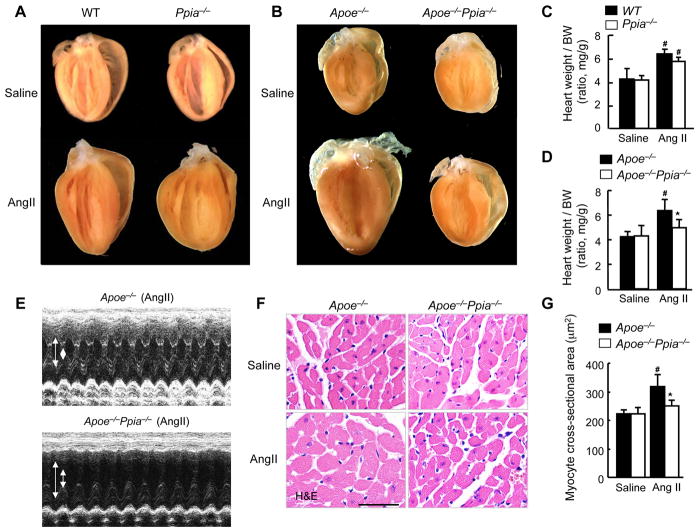
Objective: Cyclophilin A (CyPA, encoded by Ppia) is a proinflammatory protein secreted in response to oxidative stress in mice and humans. We recently demonstrated that CyPA increased angiotensin II (Ang II)-induced reactive oxygen species (ROS) production in the aortas of apolipoprotein E (Apoe)-/- mice. In this study, we sought to evaluate the role of CyPA in Ang II-induced cardiac hypertrophy.

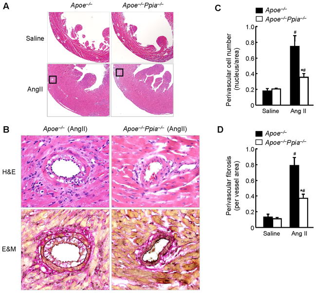
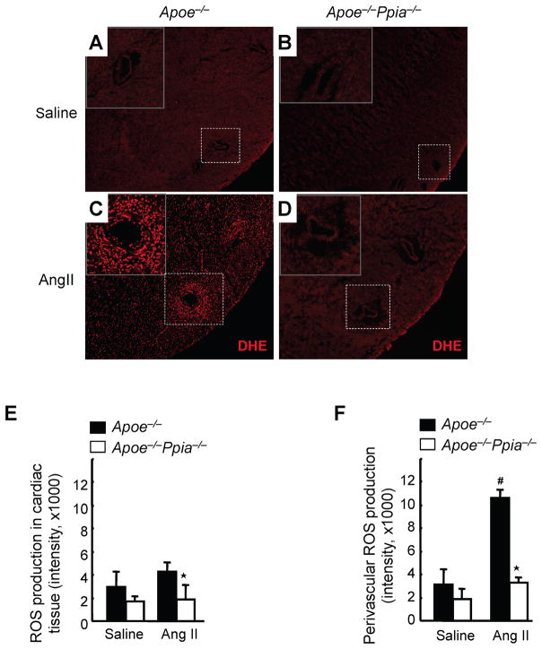
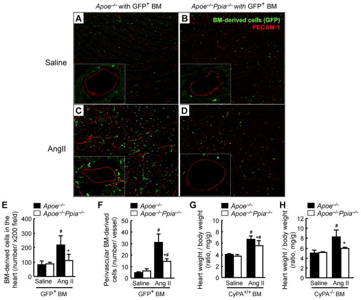
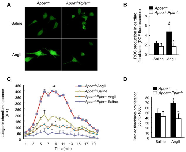
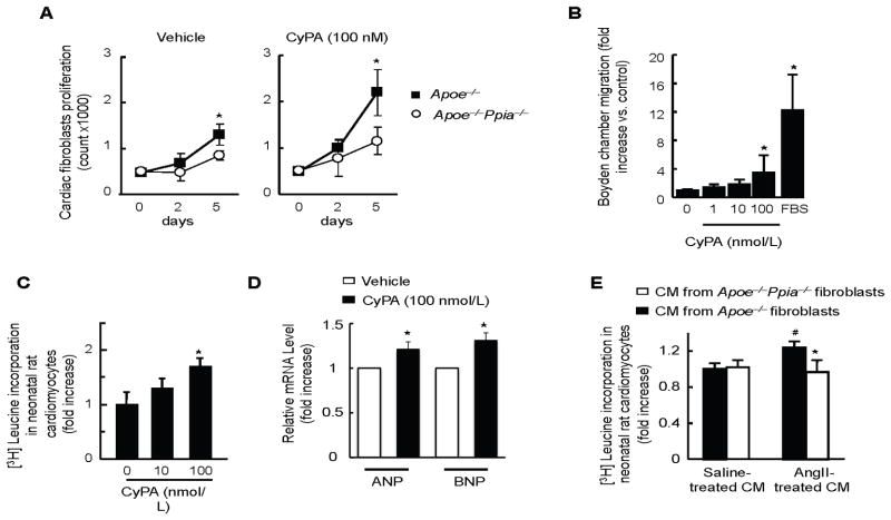
Methods and results: Cardiac hypertrophy was not significantly different between Ppia+/+ and Ppia-/- mice infused with Ang II (1000 ng/min per kg for 4 weeks). Therefore, we investigated the effect of CyPA under conditions of high ROS and inflammation using the Apoe-/- mice. In contrast to Apoe-/- mice, Apoe-/-Ppia-/- mice exhibited significantly less Ang II-induced cardiac hypertrophy. Bone marrow cell transplantation showed that CyPA in cells intrinsic to the heart plays an important role in the cardiac hypertrophic response. Ang II-induced ROS production, cardiac fibroblast proliferation, and cardiac fibroblast migration were markedly decreased in Apoe-/-Ppia-/- cardiac fibroblasts. Furthermore, CyPA directly induced the hypertrophy of cultured neonatal cardiac myocytes.

Conclusions: CyPA is required for Ang II-mediated cardiac hypertrophy by directly potentiating ROS production, stimulating the proliferation and migration of cardiac fibroblasts, and promoting cardiac myocyte hypertrophy.


Figures

Figure 1
Figure 1
CyPA deficiency prevents AngII-induced cardiac hypertrophy in Apoe−/− mice. (A) Representative photographs showing macroscopic features of cardiac hypertrophy induced by AngII infusion in Ppia−/− mice versus WT mice. AngII-induced cardiac hypertrophy is not prevented in Ppia−/− mice compared with WT mice. (B) Representative photographs of the hearts of Apoe−/− and Apoe−/− Ppia−/− mice infused with AngII or saline for 4 weeks. AngII-induced cardiac hypertrophy is prevented in Apoe−/− Ppia−/− mice compared with Apoe−/− mice. (C) AngII-induced cardiac hypertrophy (heart weight per body weight ratio, HW/BW) was not significantly decreased in Ppia−/− (n = 5) compared with WT (n = 6). # equals P< 0.05 in Saline versus AngII; *equals P< 0.05 in WT versus Ppia−/− mice. (D) AngII-induced cardiac hypertrophy (heart weight per body weight ratio) was significantly reduced in Apoe−/− Ppia−/− mice (n = 11) compared with Apoe−/− mice (n = 15). No significant differences were found in the control groups (saline infusion) of Apoe−/− and Apoe−/− Ppia−/− mice (n=4, respectively). (E) Representative M-mode images of cardiac hypertrophy assessed by echocardiography after 4 weeks of AngII infusion. Arrows show the diastolic left ventricle (LV) cavity and systolic LV cavity. (F) H&E staining of representative cross-sections of cardiac myocytes of Apoe−/− and Apoe−/− Ppia−/− mice 4 weeks after AngII infusion. (G) Myocyte cross-sectional area was significantly reduced in Apoe−/− Ppia−/− mice (n = 7) compared with Apoe−/− mice (n = 9). Results are mean ± SD. # equals P< 0.05 in Saline versus AngII; *equals P< 0.05 in Apoe−/− versus Apoe−/− Ppia−/− mice.
Figure 2
Figure 2
CyPA deficiency reduces AngII-induced perivascular cell number and fibrosis. (A) Representative H&E staining of hearts from Apoe−/− and Apoe−/− Ppia−/− mice infused with saline or AngII for 4 weeks. (B) Representative H&E and Elastica Masson (E&M) staining of coronary arteries from Apoe−/− and Apoe−/− Ppia−/− mice infused with AngII for 4 weeks. Elastic fibers stain black and collagen fibers stain red. (C) Statistical analysis of the number of cells in the perivascular area in Apoe−/− (n = 9) and Apoe−/− Ppia−/− (n = 7) mice. (D) Statistical analysis of the perivascular fibrotic area per total vascular area in Apoe−/− (n = 9) and Apoe−/− Ppia−/− (n = 7) mice. Results are mean ± SD. # equals P< 0.05 in Saline versus AngII; *equals P< 0.05 in Apoe−/− versus Apoe−/− Ppia−/− mice.
Figure 3
Figure 3
CyPA is crucial for cardiac ROS formation. (AD) Representative in situ dihydroethidium (DHE) staining of hearts. The hearts from Apoe−/− and Apoe−/− Ppia−/− mice infused with saline or AngII for 7 days were used for analysis. Images were obtained using the same magnification (x 100) and shutter speed. (EF) Densitometric analysis of oxy-ethidium (red fluorescence) in the cardiac tissue (E) and the perivascular area (F). Results are mean ± SD (n = 4 in each group). # equals P< 0.05 in Saline versus AngII; *equals P< 0.05 in Apoe−/− versus Apoe−/− Ppia−/− mice.
Figure 4
Figure 4
Bone marrow reconstitution shows a strong effect of cardiac CyPA for recruitment of bone marrow-derived cells in response to AngII, and development of cardiac hypertrophy. GFP+ bone marrow cells (Ppia+/+) were transplanted into irradiated Apoe−/− or Apoe−/− Ppia−/− mice. After 6 weeks, these chimeric mice with GFP+ bone marrow were infused with saline (AB) or AngII for 4 weeks (CD). (AD) Representative PECAM-1 staining (red) of hearts from Apoe−/− and Apoe−/− Ppia−/− recipient mice with GFP+ bone marrow (green). Statistical analysis of the number of migrating GFP+ bone marrow in the cardiac tissue (E) or perivascular area (F) in the hearts of Apoe−/− (n = 9) and Apoe−/− Ppia−/− (n = 8) mice. (G) AngII-induced cardiac hypertrophy (heart weight per body weight ratio) was significantly less in Apoe−/− Ppia−/− recipient mice (n = 9) compared with Apoe−/− recipient mice (n = 8) with Ppia+/+ bone marrow. (H) Heart weight per body weight ratio after AngII infusion for 4 weeks, was significantly less in ApoE−/− Ppia−/− (n = 6) compared to ApoE−/− (n = 6) chimeric mice with Ppia−/− bone marrow. Results are mean ± SD. # equals P< 0.05 in Saline versus AngII; *equals P< 0.05 in Apoe−/− versus Apoe−/− Ppia−/− mice.
Figure 5
Figure 5
CyPA activates cardiac fibroblasts by enhancing ROS production. (A) Representative DCF staining of mouse cardiac fibroblasts. AngII-induced ROS generation after 4 hours was decreased in CyPA-deficient cardiac fibroblasts. (B) Densitometric analysis of DCF fluorescence in response to AngII shows significant reduction in Ppia−/− cardiac fibroblasts at 4 hours (n = 8 in each group). (C) Superoxide production in cardiac fibroblasts exposed to lucingenin for 4 hours. Results are mean ± SD of three independent experiments performed in triplicate. # equals P< 0.05 in Saline versus AngII; *equals P< 0.05 in Apoe−/− versus Apoe−/− Ppia−/− mice. (D) Proliferation of cardiac fibroblasts. Apoe−/− and Apoe−/− Ppia−/− fibroblasts were treated with saline or AngII. After 48 hours of incubation, cells were counted (n = 3 in each group). Results are mean ± SD. # equals P< 0.05 in Saline versus AngII; *equals P< 0.05 in Apoe−/− versus Apoe−/− Ppia−/− cardiac fibroblasts.
Figure 6
Figure 6
CyPA promotes proliferation and migration of cardiac fibroblasts and hypertrophy of cardiac myocytes. (A) After starvation for 24 hours, cardiac fibroblasts from Apoe−/− and Apoe−/− Ppia−/− mice were stimulated with 100 nmol/L CyPA or vehicle for 5 days. Medium was changed at day 2 and cells were counted at day 2 and day 5. Data are mean ± SD. *P<0.01. n = 4 in each group. (B) Recombinant CyPA promotes cardiac fibroblasts migration in a dose-dependent manner. Data are mean ± SD. *P<0.01 compared with control. n = 6 in each group. (CD) Neonatal rat cardiac myocytes were treated with recombinant CyPA (0, 10, 100 nmol/L) for 24 hours. Hypertrophy was assessed by [3H]-leucine incorporation method (C) and by measuring ANP and BNP mRNA levels (D). Results are mean ± SD. *P < 0.05. (E) Cardiac myocytes were stimulated with conditioned medium (CM) prepared from cardiac fibroblasts that were treated with saline or AngII for 12 hours. Hypertrophy of neonatal rat cardiac myocytes was determined by means of [3H]-leucine incorporation. Data were normalized to myocytes stimulated by CM prepared from saline-treated Apoe−/− fibroblasts. n = 9 in each group. Results are mean ± SD. # equals P< 0.05 in Saline treated CM versus AngII; *equals P< 0.05 in Apoe−/− versus Apoe−/− Ppia−/− CM.

References

    1. Izumo S, Aoki H. Calcineurin--the missing link in cardiac hypertrophy. Nat Med. 1998;4:661–662. - PubMed
    1. Kakkar R, Lee RT. Intramyocardial fibroblast myocyte communication. Circ Res. 2010;106:47–57. - PMC - PubMed
    1. Souders CA, Bowers SL, Baudino TA. Cardiac fibroblast: the renaissance cell. Circ Res. 2009;105:1164–1176. - PMC - PubMed
    1. Bowers SL, Borg TK, Baudino TA. The dynamics of fibroblast-myocyte-capillary interactions in the heart. Ann N Y Acad Sci. 2010;1188:143–152. - PMC - PubMed
    1. Jaffre F, Bonnin P, Callebert J, Debbabi H, Setola V, Doly S, Monassier L, Mettauer B, Blaxall BC, Launay JM, Maroteaux L. Serotonin and angiotensin receptors in cardiac fibroblasts coregulate adrenergic-dependent cardiac hypertrophy. Circ Res. 2009;104:113–123. - PubMed

Publication types

MeSH terms