Comparative Study
. 2011 Jun;19(6):1109-17.
doi: 10.1038/oby.2011.18. Epub 2011 Feb 17.

Cafeteria diet is a robust model of human metabolic syndrome with liver and adipose inflammation: comparison to high-fat diet

Affiliations
Comparative Study

Cafeteria diet is a robust model of human metabolic syndrome with liver and adipose inflammation: comparison to high-fat diet

Brante P Sampey et al. Obesity (Silver Spring). 2011 Jun.

Abstract

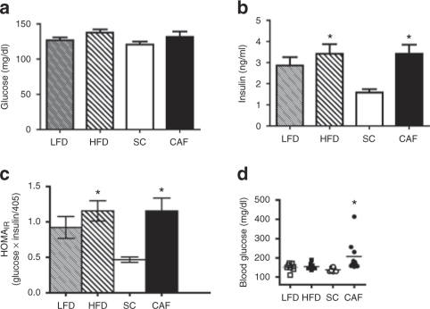
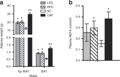
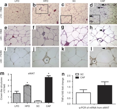
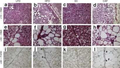
Obesity has reached epidemic proportions worldwide and reports estimate that American children consume up to 25% of calories from snacks. Several animal models of obesity exist, but studies are lacking that compare high-fat diets (HFD) traditionally used in rodent models of diet-induced obesity (DIO) to diets consisting of food regularly consumed by humans, including high-salt, high-fat, low-fiber, energy dense foods such as cookies, chips, and processed meats. To investigate the obesogenic and inflammatory consequences of a cafeteria diet (CAF) compared to a lard-based 45% HFD in rodent models, male Wistar rats were fed HFD, CAF or chow control diets for 15 weeks. Body weight increased dramatically and remained significantly elevated in CAF-fed rats compared to all other diets. Glucose- and insulin-tolerance tests revealed that hyperinsulinemia, hyperglycemia, and glucose intolerance were exaggerated in the CAF-fed rats compared to controls and HFD-fed rats. It is well-established that macrophages infiltrate metabolic tissues at the onset of weight gain and directly contribute to inflammation, insulin resistance, and obesity. Although both high fat diets resulted in increased adiposity and hepatosteatosis, CAF-fed rats displayed remarkable inflammation in white fat, brown fat and liver compared to HFD and controls. In sum, the CAF provided a robust model of human metabolic syndrome compared to traditional lard-based HFD, creating a phenotype of exaggerated obesity with glucose intolerance and inflammation. This model provides a unique platform to study the biochemical, genomic and physiological mechanisms of obesity and obesity-related disease states that are pandemic in western civilization today.


Figures

Figure 1
Figure 1
Cafeteria diet (CAF) drives food and fat consumption. (a) Male Wistar rats on CAF have elevated food intake in terms of kcal compared to standard chow (SC) control, high-fat diet (HFD), and low-fat diet (LFD). (*HFD P < 0.05 vs. all; **CAF P < 0.0001 vs. all). (b) Upon initiation of diets, only HFD-fed rats decreased food intake in grams (g) with an acute increase in CAF-intake. (*CAF P < 0.005 vs. all; ** CAF P < 0.005 vs. SC and HF; ^HF P < 0.005 vs. all; ^^HF P < 0.005 vs. SC and CAF). (c) CAF-fed rats consume more fat when compared to animals receiving HFD, LFD, or SC. (*CAF and HF P < 0.001 vs. SC and LF; ^CAF P < 0.05 vs. HF). (d) CAF-diet fed rats exhibited significant weight gain starting at 2 weeks and persisting throughout the study. Data are normalized to SC. (*CAF, LFD, or HFD P < 0.05 vs. SC, **CAF P < 0.05 vs. SC). (e) After 15 weeks on diet, CAF-fed rats exhibited greater total weight gain when compared to SC, LFD, and HFD. Although HFD-fed rats also gained weight, there is no difference in the weight gain between the HFD and LFD groups, however weight gain did reach statistical significance when compared to the SC control group. (*HF P < 0.05 vs. SC; **CAF P < 0.05 vs. all). (f) There were no changes in body length after 15 weeks on diet. (ad) were conducted on a set of rats fed diet for 10 weeks. (n = 23–24, SC, n = 8 LFD, HFD, n = 17 CAF) and (e,f) were conducted on a group of rats fed diet for 15 weeks (n = 8, SC, LFD, and HFD, n = 10 CAF).
Figure 2
Figure 2
Cafeteria diet (CAF)-fed rats displayed hyperglycemia and hyperinsulinemia. Insulin sensitivity was evaluated on rats at weeks 7 and 11 during the 15-week study. (a,b) Fasting glucose levels were not different at 7 weeks of age while only standard chow (SC)-fed rats maintained low insulin levels, which were significantly different than CAF- and high-fat diet (HFD)-fed rats. (c) Homeostasis model assessment of insulin resistance (HOMAIR) calculations at 7 weeks on diets indicated that CAF and HF groups demonstrated statistically significant elevations in insulin resistance compared to SC-fed rats. (*P < 0.05 vs. SC). (d) CAF-fed rats had significantly elevated blood glucose after a 6 h light cycle fast at 15 weeks on diet (time of sacrifice) compared to SC, while low-fat diet (LFD) and HFD maintained normal glucose levels. (n = 8, SC, LFD, and HFD, n = 10 CAF). (*P < 0.05 SC to CAF).
Figure 3
Figure 3
Cafeteria diet (CAF)-fed rats became glucose intolerant. (a) Rats fed low-fat diet (LFD) and high-fat diet (HFD) maintained similar tolerance to glucose when challenged with intraperitoneal glucose tolerance tests (IP-GTT) at 7 weeks on diets. (b) Area under the curve (AUC) quantifications for IP-GTT of averaged individual animals showed CAF-fed rats developed significant glucose intolerance compared to the standard chow (SC)-fed controls. (*P < 0.05 vs. SC; **P < 0.05 vs. SC and LF). (c) Insulin levels measured during IP-GTT at 11 weeks on diets revealed that all animals induced insulin secretion in response to glucose injections, however, SC-fed rats had lower basal insulin levels and maintained low insulin levels during the IP-GTT when compared to the other diet exposed groups. (*P < 0.05 CAF and HF vs. SC; **P < 0.05 all vs. SC; n = 8, SC, LFD, and HFD, n = 10 CAF). (d) Intraperitoneal insulin-tolerance tests (IP-ITT) revealed blunted insulin action in CAF-fed rats challenged with 0.7 IU/kg insulin when compared to SC-fed rats, but there were no significant differences between diets. HFD-fed and LFD control-fed rats displayed an intermediate insulin response that did not vary significantly between these groups. (e) AUC quantifications for IP-ITT of individual animals averaged. (n = 4, SC, LFD, and HFD, n = 5 CAF).
Figure 4
Figure 4
Dramatic increases in adiposity and nonesterified fatty acids (NEFAs) in cafeteria diet (CAF)-fed rats. (a) High-fat diet (HFD)-fed rats did not display differences in epididymal white adipose tissue (eWAT) or brown adipose tissue (BAT) size when compared to low-fat diet (LFD)-fed rats, but mass of both tissues was increased above standard chow (SC). CAF-fed animals had significantly larger eWAT and BAT fat pads compared to all groups (*P < 0.05 LF and HF vs. SC and CAF; **P < 0.05 CAF vs. all; n = 7–8, SC, LFD, and HFD, n = 10 CAF). (b) Similarly, CAF-fed rats displayed elevated circulating NEFA, with HFD and LFD groups displaying an intermediate level that were not statistically different between each other. (*P < 0.05 vs. SC; n = 4, SC, LFD, and HFD, n = 5 CAF).
Figure 5
Figure 5
Epididymal white adipose tissue (eWAT) reveals dramatic macrophage infiltration and inflammation in cafeteria diet (CAF)-fed rodents. (ah) ×10 and ×20 hematoxylin and eosin (H&E) stain of eWAT from (a,e) low-fat diet (LFD), (b,f) high-fat diet (HFD), (c,g) standard chow (SC), and (d,h) CAF. The box indicates areas of interest magnified to ×20 imaging when appropriate. Asterisks indicate crown-like structures (CLS), or areas of macrophage infiltration around adipocytes. Arrow indicates inflammatory loci. (il) Macrophage marker F4/80 was used to identify CLS (*) and areas of cells aggregating within inflammatory loci (arrow) (×40). (m) Quantification of CLS revealed a significant twofold increase in HFD- vs. LFD-fed adipose whereas CAF-fed adipose displayed a 17-fold increase over SC. (*CAF P < 0.05 vs. all, ^HFD P < 0.005 vs. all) (n) Pooled stromovascular fractions (SVF) from eWAT enriched in adipose-associated macrophages demonstrated higher expression of tumor necrosis factor α (TNFα) normalized to 18S in CAF-fed rats when compared to SC control animals as determined by qPCR. Representative images shown. (n = 4, SC, LFD, and HFD, n = 5 CAF). See Supplementary Table S4 online for detailed description.
Figure 6
Figure 6
Brown adipose tissue (BAT) macrophage infiltration and lipid droplet accumulation is perivascular. (ah) ×10 and ×40 hematoxylin and eosin (H&E) stain of BAT from (a,e) low-fat diet (LFD), (b,f) high-fat diet (HFD), (c,g) standard chow (SC), and (d,h) cafeteria diet (CAF). The box indicates area of the ×10 image magnified to ×40 imaging when appropriate for H&E stained tissues. Lipid droplet accumulation was notable in cells along the length of vessels in LFD, HFD, and especially CAF-fed tissues. (il) F4/80 was used to identify macrophages which can be seen as cells staining dark brown (marked with #) in (j) HFD and (l) CAF (×40). It is important to note that F4/80 positive macrophages colocalized to the perivascular regions where high lipid accumulation was noted and that CAF F4/80 positive cells stained stronger and were more numerous than those in HFD animals. Representative images are shown. (n = 4, SC, LFD, and HFD, n = 5 CAF). See Supplementary Table S4 online for detailed description.
Figure 7
Figure 7
Histologic analysis of rat livers demonstrates dramatic macrophage infiltration and steatohepatitis in cafeteria diet (CAF)-fed rodents. (al) ×10 and ×40 hematoxylin and eosin (H&E) stain of liver from (a,e,i) low-fat diet (LFD), (b,f,j) high-fat diet (HFD), (c,g,k) standard chow (SC), and (d,h,l) CAF. Boxes indicate regions magnified to ×40 imaging when appropriate and arrows point to inflammatory loci. Central veins (CV; eh) and portal triads (PT; il) are marked. Note healthy hepatic cord structure in (c) SC-fed livers when compared to macrovessicular steatosis in (i) LFD-fed livers and microvessicular steatotic livers in (j,l) HFD- and CAF-fed rodents. Lipid accumulation in LFD and HFD livers is localized to regions surrounding the (i,j) PT, leaving the (e,f) CV architecture intact, whereas in CAF-fed livers, lipid accumulation is (h,l) panlobular and the (d,h) hepatic cord architecture is disturbed. (mp) Serial sections relating to images il which are stained using macrophage marker F4/80 identify areas of cells aggregating into inflammatory loci (arrows) around the PT in (n) HFD livers and (p) CAF livers (×40). Representative images are shown. (q) Quantification of inflammatory loci in livers revealed a significant 1.5-fold increase in HFD- vs. LFD-fed livers while CAF-fed livers displayed more than fivefold increase (*CAF and HFD P < 0.05 vs. SC). Data are presented as the mean ± s.e.m. (n = 4, SC, LFD, and HFD, n = 5 CAF). See Supplementary Table S4 online for detailed description.

References

    1. Flegal KM, Carroll MD, Ogden CL, Curtin LR. Prevalence and trends in obesity among US adults, 1999-2008. JAMA. 2010;303:235–241. - PubMed
    1. Ogden CL, Carroll MD, Curtin LR, Lamb MM, Flegal KM. Prevalence of high body mass index in US children and adolescents, 2007-2008. JAMA. 2010;303:242–249. - PubMed
    1. Klein JD, Dietz W. Childhood obesity: the new tobacco. Health Aff (Millwood) 2010;29:388–392. - PubMed
    1. Kennedy AJ, Ellacott KL, King VL, Hasty AH. Mouse models of the metabolic syndrome. Dis Model Mech. 2010;3:156–166. - PMC - PubMed
    1. Rothwell NJ, Stock MJ. Combined effects of cafeteria and tube-feeding on energy balance in the rat. Proc Nutr Soc. 1979;38:5A. - PubMed

Publication types

MeSH terms