. 2011 Aug;18(8):1376-86.
doi: 10.1038/cdd.2011.10. Epub 2011 Feb 18.

Molecular determinants of Smac mimetic induced degradation of cIAP1 and cIAP2

Affiliations

Molecular determinants of Smac mimetic induced degradation of cIAP1 and cIAP2

M Darding et al. Cell Death Differ. 2011 Aug.

Abstract

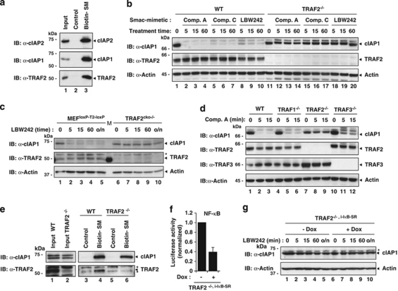
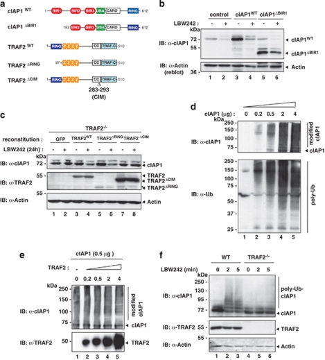
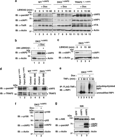
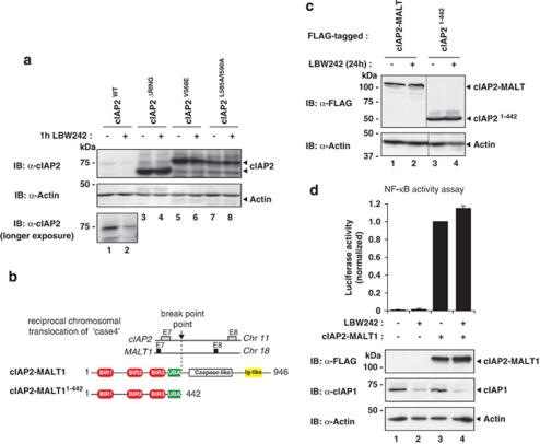
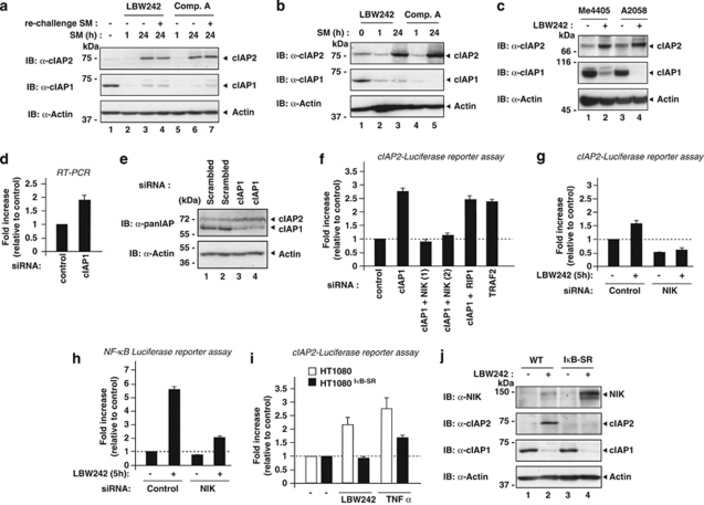
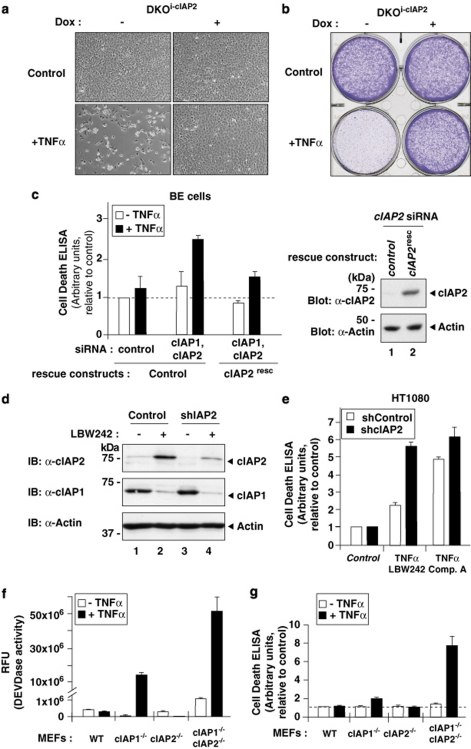
The inhibitors of apoptosis (IAP) proteins cIAP1 and cIAP2 have recently emerged as key ubiquitin-E3 ligases regulating innate immunity and cell survival. Much of our knowledge of these IAPs stems from studies using pharmacological inhibitors of IAPs, dubbed Smac mimetics (SMs). Although SMs stimulate auto-ubiquitylation and degradation of cIAPs, little is known about the molecular determinants through which SMs activate the E3 activities of cIAPs. In this study, we find that SM-induced rapid degradation of cIAPs requires binding to tumour necrosis factor (TNF) receptor-associated factor 2 (TRAF2). Moreover, our data reveal an unexpected difference between cIAP1 and cIAP2. Although SM-induced degradation of cIAP1 does not require cIAP2, degradation of cIAP2 critically depends on the presence of cIAP1. In addition, degradation of cIAP2 also requires the ability of the cIAP2 RING finger to dimerise and to bind to E2s. This has important implications because SM-mediated degradation of cIAP1 causes non-canonical activation of NF-κB, which results in the induction of cIAP2 gene expression. In the absence of cIAP1, de novo synthesised cIAP2 is resistant to the SM and suppresses TNFα killing. Furthermore, the cIAP2-MALT1 oncogene, which lacks cIAP2's RING, is resistant to SM treatment. The identification of mechanisms through which cancer cells resist SM treatment will help to improve combination therapies aimed at enhancing treatment response.


Figures

Figure 1
Figure 1
TRAF2 is required for Smac mimetic (SM)-induced cIAP1 degradation. (a and e) Biotinylated SM was used to purify SM-binding proteins from lysates of MDA-MB-231 (a) and MEFs (e). The presence of co-purified proteins was established by immunoblotting the eluate with the indicated antibodies. (bd and g) WT and TRAF-knockout MEFs were treated with 100 nM Comp. A, 100 nM Comp. C and 1 μM LBW242 for the indicated time points (min). The presence of the indicated proteins was established by immunoblotting the lysates with the indicated antibodies. Asterisks indicate nonspecific bands. (f) Secreted luciferase reporter assays using TRAF2−/−,i−IκB−SR MEFs. Cells were left untreated or treated with 100 ng/ml doxocycline (Dox) to induce IκB-SR. The medium was analysed for luciferase activity 24 h later. Luciferase activity is shown relative to the uninduced condition. The error bar indicates S.D. of triplicate experiments. (g) TRAF2−/−,i−IκB−SR MEFs were left uninduced or induced with Dox and treated with 1 μM LBW242 for the indicated time points
Figure 2
Figure 2
SM-induced cIAP1 degradation requires binding to TRAF2. (a) Schematic representation of the constructs used in b and c. (b) HEK293T cells were transiently transfected with a control plasmid or plasmids expressing either cIAP1WT or cIAP1ΔBIR1. Cells were left untreated or treated with 1 μM LBW242 for 30 min. (c) TRAF2−/− MEFs were stably infected with plasmids inducibly expressing GFP, TRAF2WT, TRAF2ΔRING and TRAF2ΔCIM, respectively. Expression of the indicated proteins was induced with 2 nM 4-hydroxy-tamoxifen, and cells were left untreated or treated with 1 μM LBW242 for 16 h before analysis. Asterisks indicate nonspecific bands. (d and e) Recombinant cIAP1 was analysed for its E3 ligase activity in vitro. The indicated amounts of cIAP1 and TRAF2 were incubated with a reaction mixture containing Ub, ATP, E1 and E2 (UbcH5c) enzymes. (f) WT and TRAF2−/− MEFs were treated with 1 μM LBW242 for the indicated time points
Figure 3
Figure 3
SM-induced degradation of cIAP2 depends on both cIAP1 and TRAF2. (a) WT, TRAF2−/− and cIAP1/2 double-knockout (DKO) MEFs stably infected with plasmids inducibly expressing cIAP2 (i-cIAP2) were treated with Dox for 16 h and exposed to 1 μM LBW242 for the indicated time points. Asterisks indicate nonspecific bands. (b) LBW242 also fails to degrade cIAP2 at later time points (24 h). (c) DKOi−cIAP1 MEFs that inducibly express cIAP1 were used to assess whether SMs stimulate cIAP1 degradation in the absence of cIAP2. cIAP1 expression was induced with 10 nM 4-hydroxy-tamoxifen for 16 h, followed by treatment with 1 μM LBW242 for the indicated time points. (df) cIAP2 expression was induced with Dox for 16 h after which cells were analysed. (d) Biotinylated SM was used to purify SM-binding proteins from lysates of the indicated MEFs. Asterisks indicate nonspecific bands. (e) cIAP2 readily promotes ubiquitylation of RIP1 in complex-I after TNFα stimulation. DKOi−cIAP1 MEFs were left untreated or treated with Dox to induce cIAP2 expression, and complex-I was purified using FLAG-tagged TNFα. Co-purified proteins were analysed by immunoblotting. (f) Induced expression of cIAP2 results in suppression of p100 cleavage. (g) cIAP1 and cIAP2 are capable of targeting NIK for degradation. NIK was co-transfected with GFP, cIAP1 or cIAP2 in 293T cells
Figure 4
Figure 4
SMs fail to stimulate degradation of cIAP2-MALT1. (a) Wild-type cIAP2, but not the RING-finger mutants cIAP2ΔRING, cIAP2V568E and cIAP2L585A/I590A, is degraded by LBW242. cIAP2ΔRING lacks the C-terminal RING-finger domain, whereas cIAP2V568E and cIAP2L585A/I590A carry point mutations that abrogate RING dimerisation and E2 binding, respectively. HEK293T cells were transiently transfected with plasmids expressing the indicated constructs. After 24 h, cells were either left untreated or incubated with 1 μM LBW242. (b) Schematic representation of the constructs used. (c) HEK293T cells were transiently transfected with plasmids expressing FLAG-cIAP2-MALT1 or FLAG-cIAP2N−Term (amino acids 1–442). The experiment was conducted as described in a. (d) Expression of cIAP2-MALT1 induces NF-κB activation that is resistant to SM intervention. cIAP2-MALT1 was co-transfected with a NF-κB reporter plasmid in HEK293T cells. Luciferase activity was expressed relative to untreated controls. Data represent the mean of three independent experiments, and the error bars indicate S.D. of triplicates
Figure 5
Figure 5
Treatment with SMs results in the upregulation of cIAP2 by non-canonical activation of NF-κB. (ac) cIAP2 protein levels increase after prolonged treatment with the SM. (a) HT1080 cells were incubated with 1 μM LBW242 or 100 nM Comp. A for the indicated time points. One hour before harvesting and lysing cells, fresh SM compounds were added (re-challenged) in the indicated lanes (LBW242 in lane and Comp. A in lane). (b) MDA-MB-231 cells were treated with 1 μM LBW242 or 100 nM of Comp. A for the indicated time points. (c) Me4405 and A2058 cells were treated for 16 h with 100 nM of Comp. A. (d) Cells were transfected with the indicated siRNA oligos, harvested and lysed 2 days after transfection. (e) cIAP2 mRNA levels increase upon cIAP1 depletion. BE cells were transfected with the indicated siRNA oligos and analysed after 48 h using quantitative RT-PCR. (f and g) SM treatment and siRNA-mediated depletion of cIAP1 and TRAF2 result in the activation of non-canonical NFκB signalling and induction of cIAP2 gene expression in BE cells. Cells stably expressing a luciferase reporter under the control of the cIAP2 promoter (cIAP2-Luc) were transfected with the indicated siRNA oligos and analysed for luciferase activity 48 h later. (h) SM treatment results in NIK-dependent activation of NF-κB. HT1080 cells stably expressing a NF-κB luciferase reporter were transfected with the indicated siRNA oligos and analysed for luciferase activity 48 h later. (i and j) SM- and TNFα-mediated induction of cIAP2 expression is NF-κB dependent. (i) HT1080 and HT1080IκB−SR cells, which express a non-degradable form of IκB, were transfected with the cIAP2-Luc reporter plasmid and left untreated or treated with the indicated combinations. (j) Inhibition of SM-mediated induction of cIAP2 results in elevated levels of NIK. HT1080 and HT1080IκB−SR cells were left untreated or treated with 1 μM LBW242 for 16 h
Figure 6
Figure 6
Induction of cIAP2 provides resistance to SM-induced TNFα killing in cancer cells. (a and b) Induced expression of cIAP2 protects cells against TNFα-mediated cell death in the absence of cIAP1. (a) Light micrographs showing the overall morphology of DKOi−cIAP2 MEFs treated with the indicated combinations. (b) Clonogenic survival assay. Cells were visualised using crystal violet. (c) BE cells, stably expressing either a control plasmid or siRNA-resistant cIAP2 (cIAP2resc), were transfected with the indicated siRNA oligos. After 48 h, cells were left untreated or treated with TNFα for 18 h. Cell death was assayed using Cell Death ELISA (Roche Applied Science, Penzburg, Germany) (measuring the amount of cleaved DNA–histone complexes (nucleosomes) in the cytoplasm; left panel). cIAP2 expression of the RNAi-resistant rescue construct was examined by immunoblotting the lysates with an anti-cIAP2 antibody (right panel, lane 2). (d and e) RNAi-mediated knockdown of cIAP2 increases sensitivity to TNFα killing upon SM treatment. HT1080 cells stably expressing either a control shRNA or shcIAP2 were either left untreated or treated with SM for 16 h. (d) The expression of cIAP2 was analysed by immunoblotting. (e) Cell Death ELISA of HT1080shControl and HT1080shcIAP2 cells treated with the indicated combinations. (f and g) cIAP1/2 DKO MEFs are significantly more sensitive to TNFα than singly cIAP1−/− MEFs. The indicated MEFs were exposed to TNFα, and caspase activity and apoptosis were determined using DEVDase assays (f) and Cell Death ELISA (g)

References

    1. Chen DJ, Huerta S. Smac mimetics as new cancer therapeutics. Anticancer Drugs. 2009;20:646–658. - PubMed
    1. Vaux DL, Silke J. Mammalian mitochondrial IAP binding proteins. Biochem Biophys Res Commun. 2003;304:499–504. - PubMed
    1. Cossu F, Mastrangelo E, Milani M, Sorrentino G, Lecis D, Delia D, et al. Designing Smac-mimetics as antagonists of XIAP, cIAP1, and cIAP2. Biochem Biophys Res Commun. 2009;378:162–167. - PubMed
    1. Yang YL, Li XM. The IAP family: endogenous caspase inhibitors with multiple biological activities. Cell Res. 2000;10:169–177. - PubMed
    1. Gyrd-Hansen M, Darding M, Miasari M, Santoro MM, Zender L, Xue W, et al. IAPs contain an evolutionarily conserved ubiquitin-binding domain that regulates NF-∣[kappa]∣B as well as cell survival and oncogenesis. Nat Cell Biol. 2008;10:1309, 1317. - PMC - PubMed

Publication types

MeSH terms

Substances