Review
. 2011 Mar;12(3):154-67.
doi: 10.1038/nrn2994.

The integration of negative affect, pain and cognitive control in the cingulate cortex

Affiliations
Review

The integration of negative affect, pain and cognitive control in the cingulate cortex

Alexander J Shackman et al. Nat Rev Neurosci. 2011 Mar.

Abstract

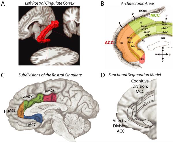
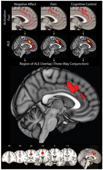
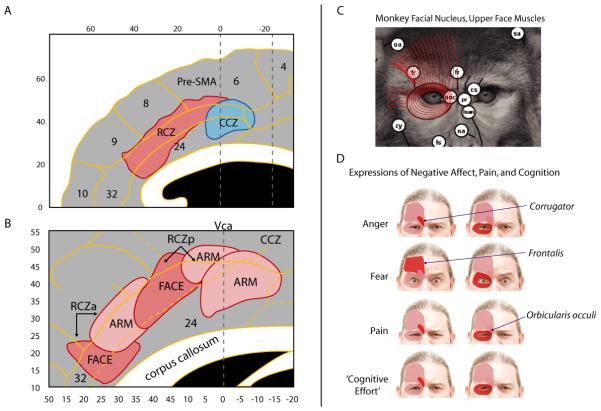
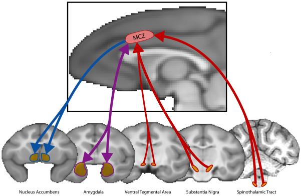
It has been argued that emotion, pain and cognitive control are functionally segregated in distinct subdivisions of the cingulate cortex. However, recent observations encourage a fundamentally different view. Imaging studies demonstrate that negative affect, pain and cognitive control activate an overlapping region of the dorsal cingulate--the anterior midcingulate cortex (aMCC). Anatomical studies reveal that the aMCC constitutes a hub where information about reinforcers can be linked to motor centres responsible for expressing affect and executing goal-directed behaviour. Computational modelling and other kinds of evidence suggest that this intimacy reflects control processes that are common to all three domains. These observations compel a reconsideration of the dorsal cingulate's contribution to negative affect and pain.


Figures

Figure 1
Figure 1. Divisions of the human rostral cingulate
The rostral cingulate has been partitioned on physiological and anatomical grounds at spatial scales ranging from the macroscopic to the molecular. A. Three-dimensional rendering of the left rostral cingulate cortex. The cingulate, shown in red, was manually traced on a single subject’s magnetic resonance image (MRI). Much of the constituent cortical gray matter lies buried within the cingulate sulci, a fact not apparent from inspection of the mesial surface (for additional information, see Box 1 and the Supplement). B. Architectonic areas of the cingulate. Areas were defined on the basis of differences in microanatomy and neurotransmitter chemistry and, hence, differ somewhat from the classical descriptions of Brodmann and other pioneering neuroanatomists, . Architectonic features provide one means of defining homologies across species, . Adapted with permission from Ref. . C. The four major subdivisions of the rostral cingulate. Subdivisions were defined by Vogt and colleagues on the basis of regional differences in microanatomy, connectivity, and physiology. The supracallosal portion of the cingulate is designated the midcingulate cortex (MCC) and is divided into anterior (aMCC: green) and posterior (pMCC: magenta) subdivisions. The portion of the cingulate lying anterior and ventral to the corpus callosum is designated the anterior cingulate cortex (ACC) and is divided into pregenual (pgACC: orange) and subgenual (sgACC: cyan) subdivisions by the coronal plane at the anterior tip of the genu. Adapted with permission from Ref. (for additional information, see the Supplement). D. The functional segregation model of Bush, Luu and Posner. On physiological and anatomical grounds, Bush et al. argued that the rostral cingulate consists of two functionally segregated regions: a rostroventral ‘affective’ division (ACC; originally termed ‘ventral ACC’) and a dorsal ‘cognitive’ division (MCC; originally termed ‘dorsal ACC’). Adapted with permission from Ref. .
Figure 2
Figure 2. Negative affect, pain, and cognitive control activate a common region within anterior midcingulate cortex (aMCC)
Map depicts the results of a coordinate-based meta-analysis (CBMA) of 380 activation foci derived from 192 experiments involving more than 3,000 participants. Uppermost row shows the spatially normalized foci for each domain. The next row shows thresholded activation likelihood estimate (ALE) , maps for each domain considered in isolation. Bottom two rows depict the region of overlap across the three domains. Red cluster indicates the location of a three-way minimum significance conjunction of the three domains. The cluster lies in aMCC in the vicinity of areas 32′ and a24b’/c’ (Talairach coordinates: x=0, y=12, z=42; volume: 11680 mm3). No other cluster reached significance. Numbers indicate mm from the anterior commissure (for additional methodological details and results, see the Supplement).
Figure 3
Figure 3. Cingulate premotor areas in the human midcingulate cortex (MCC)
A. Locations of the rostral and caudal cingulate zones (RCZ and CCZ), . RCZ lies in aMCC, whereas CCZ lies at the junction of aMCC and posterior MCC (pMCC) (see Figure 1c). Zone borders are approximations (see also Ref. 44). Adapted with permission from Ref. (for additional information, see the Supplement). B. Somatotopy in RCZ and CCZ based on human imaging studies. Adapted with permission from Ref. (for additional information, see the Supplement). C. Combined tracing and microstimulation work in macaques indicates that the monkey analogue of the human RCZ projects to the facial nucleus, , allowing it to control the muscles of the upper face (shown in red for the macaque). The facial muscles are largely conserved across primate species, . Adapted with permission from Ref. (for additional information, see the Supplement). D. In humans, the muscles of the upper face have been associated with the elicitation of negative affect (e.g., anger, fear), pain, and consistent with Darwin’s suggestions perhaps ‘cognitive effort’ as well (for additional information, see the Supplement).
Figure 4
Figure 4. Subcortical connnectivity of the macaque analogue to the human Rostral Cingulate Zone (RCZ)
Afferents are depicted in red, efferents in blue, and reciprocal connections in purple. The monkey analogue to RCZ receives substantial inputs from the spinothalamic system, which relays nociceptive information from the periphery to RCZ via the mediodorsal nucleus of the thalamus. Dopaminergic inputs to RCZ arise from the substantia nigra and, to a lesser extent, the ventral tegmental area. RCZ projects to the ventral striatum, including the core region of nucleus accumbens, and has robust reciprocal connections with the lateral basal nucleus of the amygdala. For additional information, see the Supplement.
Box 1 Figure
Box 1 Figure

References

    1. Brodmann K. The principles of comparative localisation in the cerebral cortex based on cytoarchitectonics. Springer; New York: 1909/2005. Brodmann’s: Localisation in the cerebral cortex.
    1. Papez JW. A proposed mechanism of emotion. Arch Neurol Psychiatry. 1937;38:725–733.
    1. Kober H, et al. Functional grouping and cortical-subcortical interactions in emotion: a meta-analysis of neuroimaging studies. Neuroimage. 2008;42:998–1031. - PMC - PubMed
    1. Tracey I, Mantyh PW. The cerebral signature for pain perception and its modulation. Neuron. 2007;55:377–391. - PubMed
    1. Vogt BA, Sikes RW. Cingulate nociceptive circuitry and roles in pain processing: The cingulate premotor pain model. In: Vogt BA, editor. Cingulate neurobiology and disease. Oxford University Press; NY: 2009. pp. 311–338.

Publication types