

Review
doi: 10.1038/nrn2995.
Engaging neuroscience to advance translational research in brain barrier biology
Affiliations
- PMID: 21331083
- PMCID: PMC3335275
- DOI: 10.1038/nrn2995
Item in Clipboard
Review
Engaging neuroscience to advance translational research in brain barrier biology
Nat Rev Neurosci.
2011 Mar.
Abstract
The delivery of many potentially therapeutic and diagnostic compounds to specific areas of the brain is restricted by brain barriers, of which the most well known are the blood-brain barrier (BBB) and the blood-cerebrospinal fluid (CSF) barrier. Recent studies have shown numerous additional roles of these barriers, including an involvement in neurodevelopment, in the control of cerebral blood flow, and--when barrier integrity is impaired--in the pathology of many common CNS disorders such as Alzheimer's disease, Parkinson's disease and stroke.
Figures
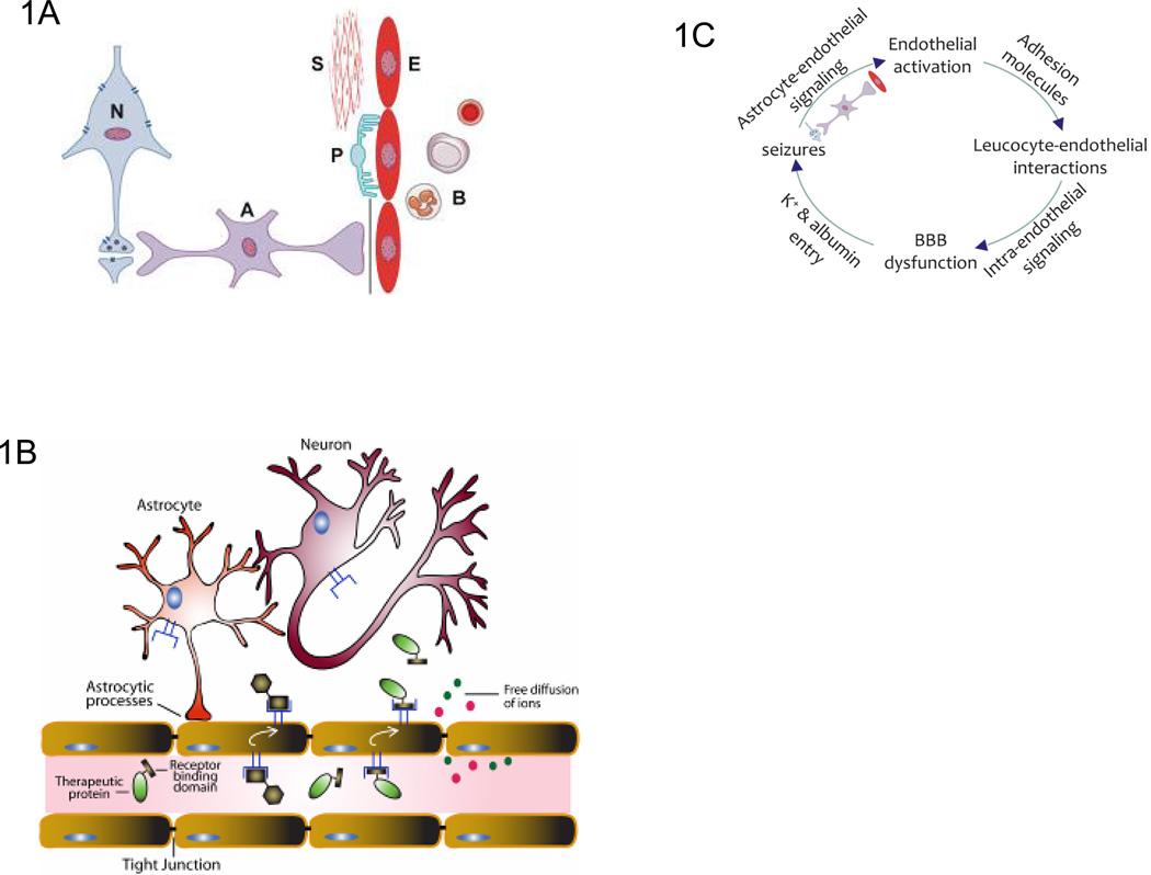
a. The blood-brain barrier (BBB) is an essential part of the neurovascular unit (NVU). A classical view of the NVU incorporates neurons, glial cells such as astrocytes and microglial cells closely juxtaposed with vascular endothelial cells, pericytes and smooth muscle cells. Blood cells, particularly PMN cells, lymphocytes and monocytes, also interact with the BBB endothelium and are therefore an integral part of this unit. The interactions between these cellular components and inter- and intra-cellular signaling regulate NVU function to maintain homeostasis, or to respond to inflammation and disease. b. Receptor-mediated transcytosis of proteins at the BBB. Transcytosis is a receptor-mediated transport mechanism by which proteins that are targeted to the CNS bind extracellular receptors in vascular lumen, transport across the BBB endothelial cells, and are released in brain parenchyma. The presence of specific receptors (i.e. the insulin receptor) on the surface of BBB endothelial cells has allowed targeting and transport of some therapeutic proteins to the CNS, . c. Pathological signaling in the extended NVU. The proposed sequence order is based on data available from the epilepsy field and requires further exploration in the context of other brain diseases, including stroke and AD. The cycle starts with altered expression of vascular cell adhesion molecules and interactions of leucocytes with the endothelium, initiating intra-endothelial signals that alter BBB function and lead to neural tissue dysfunction as a consequence of K+ and albumin entry into the brain interstitium. Astrocytes detect the altered neuronal activity and transmit signals back to the BBB thereby facilitating interactions with leucocytes and turning the sequence into a vicious circle that maintains and exacerbates the pathological state. The activated endothelium may, as an integral part of the extended NVU, disturb neuron–astrocyte interactions, thereby adding an additional layer of pathological signaling to the process. Astrocytes emerge from this cascade as a primary target for interventions that aim to interrupt the proposed cycle.
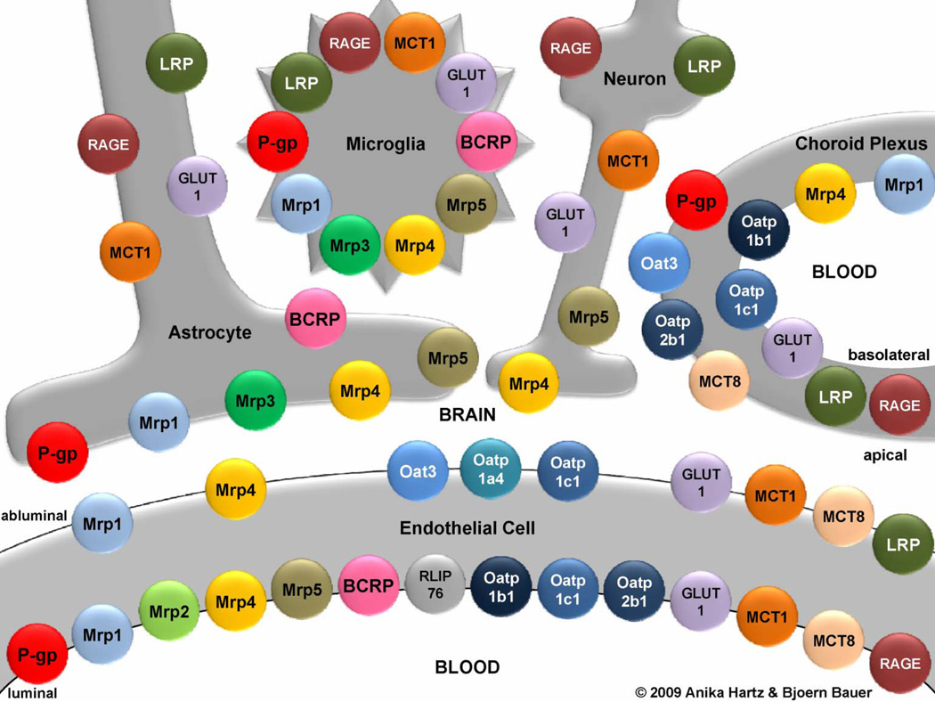
The spatial and cellular relationships of the transporters are shown. Only proteins detected at the protein level are depicted. For a more complete listing of carrier-mediated transport systems at the blood-brain interface see Ohtsuki and Terasaki. The following acronyms are used: BCRP, breast cancer resistance protein; GLUT1, glucose transporter-1; LRP, low-density lipoprotein receptor-related protein; MCT, monocarboxylic acid transporter; Mrp, multidrug resistance protein; Oat, organic anion transporter; Oatp, organic anion transporting protein; P-gp, P-glycoprotein; RAGE, receptor for advanced glycation end products; RLIP76, Ral-binding protein-1. Modified, with permissions, from Refs. and .
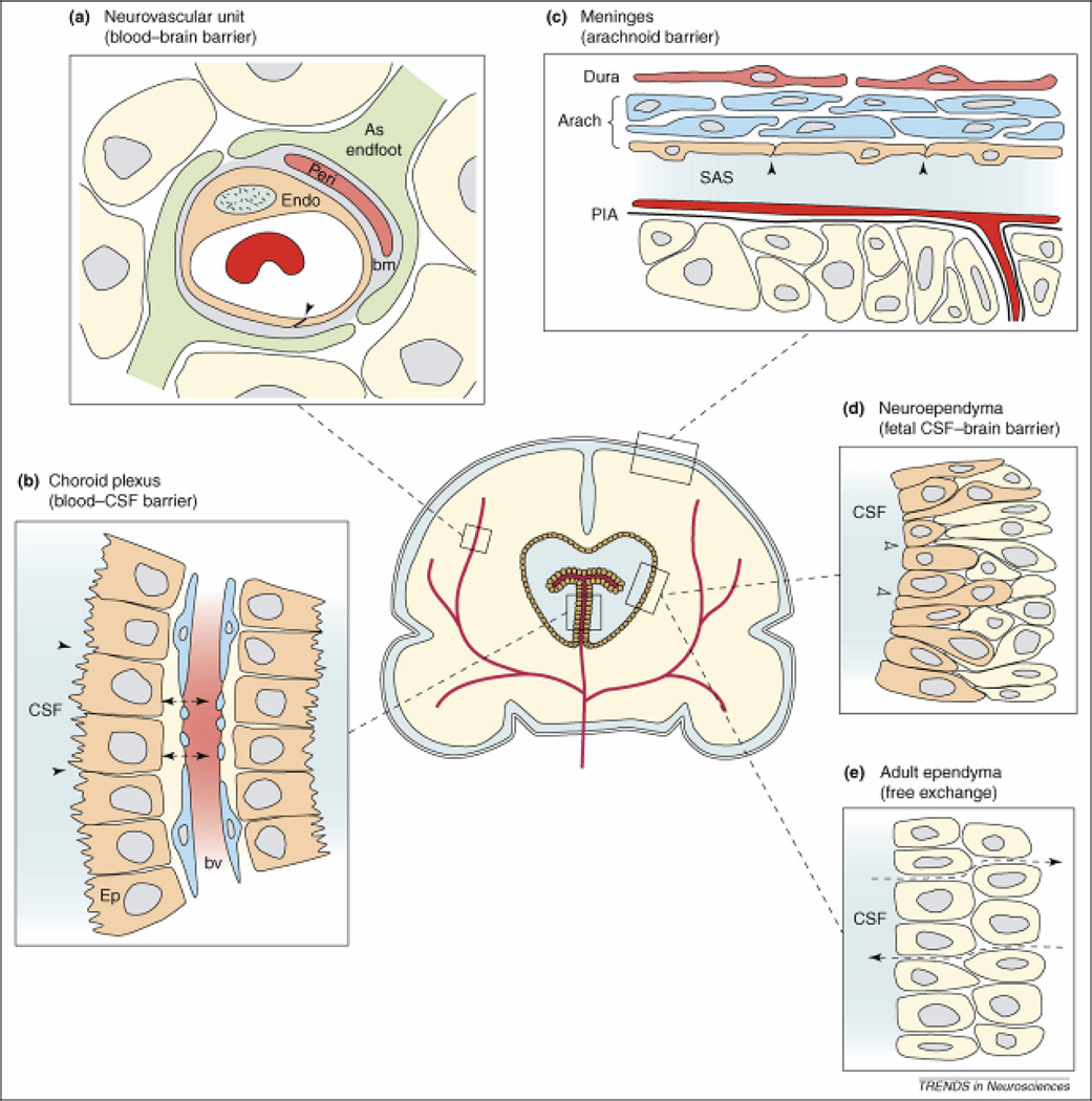
The neurovascular unit (a), blood-CSF barrier (b), and arachnoidal barrier (c) are common between developing and adult brain, whereas fetal neuroependyma (d) differs from adult ependyma (e). (a) Endothelial cells (Endo) have luminal tight junctions (arrowhead) forming the physical barrier of the interendothelial cleft. Outside the endothelial cell is a basement membrane (bm) which also surrounds the pericytes (Peri). Around all these structures are the astrocyctic endfeet processes from nearby astrocytes (As Endfoot). (b) The endothelial cells of choroid plexus blood vessels are fenestrated and form a non-restrictive barrier (arrowheads) between the cerebrospinal fluid (CSF) and blood vessel (BV). The epithelial cells (Ep) have apical tight junctions (small arrows) that restrict intercellular passage of molecules. (c) In the meninges, the blood vessels of the dura are fenestrated and provide little barrier function (not shown); however, the outer cells of the arachnoid membrane (Arach) have tight junctions (arrowheads) and this cell layer forms the physical barrier between the CSF-filled subarachnoid space (SAS) and overlying structures. The blood vessels between the arachnoid and the pial surface (PIA) have tight junctions (not shown). (d) In early development the neuroependymal cells are connected to each other by strap-junctions (small arrows) that are believed to form the physical barrier restricting the passage of larger molecules such as proteins but not smaller molecules such as sucrose. (e) The mature adult ventricular ependyma does not restrict the exchange of molecules. Reproduced, with permissions, from Ref. .
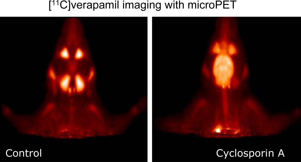
Micro PET images of the head of a Wistar rat, showing the biodistribution of the calcium channel blocker, [11C] verapamil injected systemically, either alone (Control) or after pre-treatment of the animal with the Pgp inhibitor Cyclosporin A. [11C] verapamil, a substrate for the blood-brain barrier efflux transporter P-glycoprotein, gains access to the brain only after Pgp inhibition by Cyclosporin A. Images are courtesy of Dr. P. Elsinga, University Medical Center Groningen, The Netherlands.
References
-
- Selkoe DJ. Alzheimer disease: mechanistic understanding predicts novel therapies. Ann Intern Med. 2004;140:627–638. - PubMed
-
- Zlokovic BV. Neurovascular mechanisms of Alzheimer's neurodegeneration. Trends Neurosci. 2005;28:202–208. - PubMed
-
- Kortekaas R, et al. Blood-brain barrier dysfunction in parkinsonian midbrain in vivo. Ann Neurol. 2005;57:176–179. - PubMed
-
- Gold R, Linington C, Lassmann H. Understanding pathogenesis and therapy of multiple sclerosis via animal models: 70 years of merits and culprits in experimental autoimmune encephalomyelitis research. Brain. 2006;129:1953–1971. - PubMed
Publication types
MeSH terms
Substances
Grants and funding
LinkOut - more resources
Full Text Sources
Other Literature Sources
Medical
