. 2004 Sep;21(3):205-16.
doi: 10.1055/s-2004-860941.

Colon stenting: a review

Affiliations

Colon stenting: a review

Miguel Angel de Gregorio et al. Semin Intervent Radiol. 2004 Sep.

Abstract

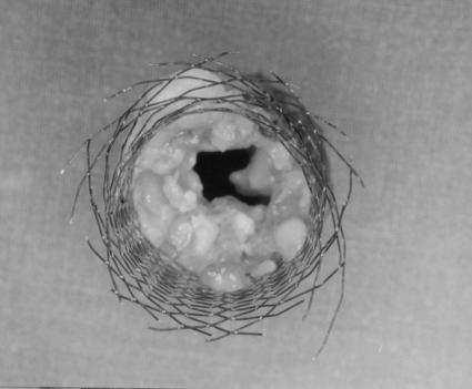
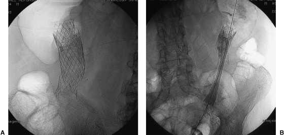
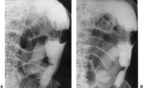
Up to 85% of patients who present with colonic obstruction have a colorectal cancer. Between 7% and 29% of these patients present with total or partial intestinal obstruction. Only 20% of these patients presenting with acute colonic obstruction due to malignancy survive 5 years. Emergent surgical intervention in patients with colonic obstruction is associated with significant morbidity and mortality rates. Only 40% of patients with obstructive carcinoma of the left colon can be treated with surgical resection without the need for a colostomy. The use of a temporary or permanent colostomy has a significant impact on quality of life. The decompressive effect seen with colonic stenting is a durable, simple, and effective palliative treatment of patients with advanced disease. Stent deployment provides an effective solution to acute colonic obstruction and allows surgical treatment of the patient in an elective and more favorable condition. In addition, colonic stenting reduces costs and avoids the need for a colostomy.

Keywords: Colon cancer; colonic stenting; intestinal obstruction.


Figures

Figure 1
Figure 1
Photograph of an enteral Wallstent. This enteral endoprosthesis is a self-expandable metallic stent designed for colonic use. It consists of a monofilament wire. It has a diameter of 18 to 22 mm and is available in lengths of 6 and 9 cm (courtesy of Meditech-Boston Scientific).
Figure 2
Figure 2
Photograph of colonic Z-stent. The colonic endoprosthesis Z-stent is a self-expandable metallic stent. It has a flared end diameter of 35 mm and a shaft diameter of 25 mm. It is available in 4- and 12-cm lengths (courtesy of Cook Europe).
Figure 3
Figure 3
Photograph of a Precision Wallstent. The Precision endoprosthesis is a self-expandable metallic stent. It has a flared end diameter of 30 mm and a shaft diameter of 25 mm. It is available in 6- and 12-cm lengths. The Precision delivery system is a 16 F sheath (courtesy of Microvasive, Boston Scientific).
Figure 4
Figure 4
Photograph of a covered colonic Choostent. The Choostent is a stainless cylindrical zigzag stent. It is available as a covered and uncovered stent (courtesy of Life Europe).
Figure 5
Figure 5
Surgical specimen from colectomy. Wallstent visualized within the tumor. Free wire filaments are appreciated in the distal portion of the stent.
Figure 6
Figure 6
Diagrams demonstrate colorectal stent deployment with fluoroscopic guidance. An obstructive carcinoma is present in the descendent colon. (A) The guide wire and the catheter are advanced through the area of obstruction. (B) The guide wire is replaced by an Amplatz stiff guide wire to straighten the tortuous colon, and the delivery system is introduced. (C) The stent is initially deployed at the proximal portion of the lesion. (D) The stent is fully expanded.
Figure 7
Figure 7
Images obtained during fluoroscopic placement of a colonic stent showing the stent deployment technique. (A) Neoplasia within the descending colon. The guide wire has crossed the area of obstruction. (B) Precison stent partially deployed in the area of malignant obstruction. The delivery system can be seen. (C) Stent totally deployed and partially expanded. (D) Radiographic control with stent totally expanded.
Figure 8
Figure 8
Lateral view of the abdomen. Colonoscope can be seen close to a lesion in the sigmoid colon.
Figure 9
Figure 9
Utilization of a 10-F introducer sheath to facilitate the technique. (A) Introduction of contrast through the sheath to determine the morphology and the extension of the lesion keeping a guide wire distal to it. (B) Gathering a biopsy sample with a forceps through the sheath, guide wire distal to the lesion for safety purposes.
Figure 10
Figure 10
Antegrade approach for the deployment of a colonic stent. (A) Guide wire crossing the area of malignant obstruction with access through a percutaneous cecostomy. (B) Delivery system introduction carrying the stent through a 10-F sheath placed in the cecostomy. (C) Radiographic control after stent placement. A 10-F pigtail catheter is present in the cecum. (D) Control performed 3 months after stent placement showing patency of the stent. Pigtail catheter was withdrawn without complications.
Figure 11
Figure 11
(A) Single view of the abdomen showing significant abdominal distention with dilatation of the colon proximal to the site of obstruction. (B) Radiographic control 24 hours postimplantation of the stent. Resolution of the obstructive pattern.
Figure 12
Figure 12
Implantation of a colonic stent with a combined guidance (fluoroscopy and colonoscopy). (A) Colonoscope is close to the area of obstruction within the sigmoid colon. (B) Endoscopic view of the area of obstruction.
Figure 13
Figure 13
Stent migration. (B) Wallstent in posteroanterior (PA) view within the rectosigmoid lesion. Remains of barium can be seen. (B) Stent migration and change of position. (C) Radiographic control after stent expulsion.
Figure 14
Figure 14
Surgical specimen from colectomy performed 24 days after stent implantation. Tumoral growth through the stent with narrowing of the lumen is noted. The patient remained asymptomatic.
Figure 15
Figure 15
Rupture of a stent. A 76-year-old patient diagnosed with and treated for colon carcinoma 7 years previously. Placement of stent to treat a left colon lesion. Stent was implanted without complications (Memotherm 25 × 80 mm). Thirty months after procedure patient presented with new intestinal obstruction. Abdominal exam showed rupture of the stent. A new stent was implanted without incident (Wallstent 24 × 70 mm). (Images provided by Dr. J. Urbano.)
Figure 16
Figure 16
Intestinal obstruction with lesion in the splenic angle. (A) Apple-core lesion seen with guide wire crossing it. (B) Image after deployment.

References

    1. Boyle P, Leon M. Epidemiology of colorectal cancer. Br Med Bull. 2002;64:1–25. - PubMed
    1. Deans G T, Krukowski Z H, Irwin S T. Malignant obstruction of the left colon. Br J Surg. 1994;81:1270–1276. - PubMed
    1. Duxbury M, Brodribb A, Oppong F, Hosie K. Management of colorectal cancer: variations in practice in one hospital. Eur J Surg Oncol. 2003;29:400–402. - PubMed
    1. Griffith R. Prospective medical obstacles to surgery. Cancer. 1992;70:1333–1341. - PubMed
    1. Smothers L, Hynan L, Fleming J, Turnage R, Simmang C, Anthony T. Emergency surgery for colon carcinoma. Dis Colon Rectum. 2003;46:24–30. - PubMed