. 2004 Dec;21(4):221-33.
doi: 10.1055/s-2004-861556.

Vascular complications following liver transplantation

Affiliations

Vascular complications following liver transplantation

James C Andrews. Semin Intervent Radiol. 2004 Dec.

Abstract

Vascular complications (stenosis or thrombosis of the hepatic artery, portal vein or hepatic vein) are a relatively common occurrence following liver transplantation. Routine screening with ultrasound is critical to early detection of these complications. Careful application of standard interventional techniques (diagnostic catheter angiography, balloon angioplasty with selective stenting) may be used to confirm the ultrasound findings, treat the underlying lesions, and contribute to long-term graft survival.

Keywords: Liver transplantation; balloon angioplasty; hepatic artery; hepatic vein; portal vein.


Figures

Figure 1
Figure 1
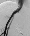
Normal celiac arteriogram demonstrating a widely patent arterial anastomosis. Note that both the donor and recipient gastroduodenal arteries have been ligated.
Figure 2
Figure 2
Anteroposterior (A) and left anterior oblique (B) aortograms demonstrate the infrarenal aorta to hepatic artery conduit. The widely patent origin is seen well only on the oblique view. A selective injection (C) into the conduit better depicts the stenosis at the distal anastomosis.
Figure 3
Figure 3
Selective injections into the aortohepatic conduit (A) and the common hepatic artery (B) from a different patient demonstrate thrombosis of the hepatic artery. Knowledge of the surgical anatomy is critical in these patients to make the diagnosis of hepatic artery thrombosis correctly.
Figure 4
Figure 4
Selective injection into the left hepatic vein in a patient with a piggy-back hepatic vein anastomosis (A). Contrast material (CO2 in this case) refluxes into all the major transplant hepatic vein branches because of an anastomotic stenosis (not well seen in this projection). This projection does give an overall view of the relationship of the hepatic veins to the recipient inferior vena cava (IVC). Selective injection into the right hepatic vein from another patient (B) demonstrates reflux into the ligated donor IVC stump. This should not be confused with a thrombosed hepatic vein branch.
Figure 5
Figure 5
Selective hepatic arteriograms before (A) and after (B) guidewire manipulation across the anastomotic stenosis. Note the diffuse vasospasm throughout the entire liver. This was only partially broken with intra-arterial nitroglycerin. At the time of repeated intervention 2 months later, intra-arterial nitroglycerin was given prophylactically before a wire was advanced across with anastomosis and no vasospasm was encountered.
Figure 6
Figure 6
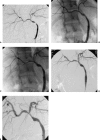
Hepatic arteriogram demonstrating a stenosis at the anastomosis between the donor and recipient hepatic arteries (A). The lesion was crossed with a 0.018-inch wire and dilated with a 4-mm balloon (B). Follow-up angiogram (C) shows a satisfactory PTA result.
Figure 7
Figure 7
Hepatic arteriogram obtained by injecting the aortohepatic artery conduit identifies a stenosis at the distal anastomosis (A). After a guide catheter was placed into the proximal portion of the conduit (B), the lesion was crossed and dilated. In this case a 6F bright-tip sheath was used as a guide. Follow-up injection through the guide catheter (C).
Figure 8
Figure 8
Initial hepatic arteriogram obtained by injecting the aortohepatic artery conduit demonstrates a recurrent stenosis at the distal anastomosis (A). There had been two prior dilatations of this lesion. Follow-up run after PTA using a 4-mm balloon (B) shows active extravasation from the ruptured hepatic artery. The balloon was reinflated to control the hemorrhage and the artery repaired operatively.
Figure 9
Figure 9
Recurrent stenosis (one prior PTA) at the distal anastomosis of a aortohepatic artery conduit (A). Follow-up angiogram after placement of a balloon-expandable stent across the anastomosis (B).
Figure 10
Figure 10
Typical stenosis at the distal anastomosis of an aortohepatic artery conduit (A). Follow-up run after PTA with a 3-mm balloon demonstrates a dissection in the donor hepatic artery (B). Additional run obtained 5 minutes later shows extension of the dissection (C). The flap was tacked down with a balloon-expandable stent (D) with a good technical and functional result. Follow-up angiogram 8 months later, obtained because of an abnormal ultrasound study, shows some intimal hyperplasia within the stent (E). Liver function remained normal.
Figure 11
Figure 11
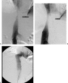
Arterial portogram of a 3-year-old patient, obtained by injecting the superior mesenteric artery (SMA) (A), identifies a portal vein stenosis with large left upper quadrant varices. Direct portogram obtained from the transhepatic approach better demonstrates the stenosis (B). Follow-up venogram after PTA shows resolution of the stenosis (C).
Figure 12
Figure 12
Transhepatic portogram shows near-complete obstruction at the portal anastomosis (A). Immediate elastic recoil following PTA (not shown) was treated by placing a balloon-expandable stent across the anastomosis (B).
Figure 13
Figure 13
This 12-year-old patient had a portal vein stenosis treated with balloon-expandable stents at age 18 months. Transhepatic portogram shows that the stents have separated (A) and are now too small for the portal vein. Multiple collaterals drain around the stents (B). No intervention was thought to be possible.
Figure 14
Figure 14
Inferior venacavagram using gadolinium as contrast agent shows a tight stenosis of the intrahepatic IVC (A). Follow-up run after PTA with an 18-mm balloon shows a patent but narrowed IVC with good flow into the right atrium (B). Cavagram in another patient following Gianturco stent placement demonstrates a widely patent IVC (C). Note the large spaces between the stent struts, which minimize obstruction to inflow from side branches such as the hepatic veins.
Figure 15
Figure 15
Left hepatic venogram in a patient with a piggy-back anastomosis. Note the reflux of contrast material into the right hepatic vein due to the anastomotic stenosis (A). This would have been more easily done from a jugular puncture, but the presence of an implanted central venous catheter precluded this approach. Venogram after PTA (B) of the anastomosis shows resolution of the stenosis. Reflux into the right hepatic vein was no longer seen.
Figure 16
Figure 16
Right hepatic venogram from the same patient as in Fig. 4B shows placement of a balloon-expandable stent to treat the anastomotic stenosis. Despite the stent crossing the origins of the left and middle hepatic veins, the patient's symptoms (ascites) resolved quickly after stent placement.

Similar articles

Cited by

References

    1. Orons P D, Zajko A B, Bron K M, et al. Hepatic artery angioplasty after liver transplantation: experience in 21 allografts. J Vasc Interv Radiol. 1995;6:523–529. - PubMed
    1. Soin A S, Friend P J, Rasmussen A, et al. Donor arterial variations in liver transplantation: management and outcome of 527 consecutive grafts. Br J Surg. 1996;83:637–641. - PubMed
    1. Neuhaus P, Platz K P. Liver transplantation: newer surgical approaches. Baillieres Clin Gastroenterol. 1994;8:481–493. - PubMed
    1. Cappadonna C R, Johnson L B, Lu A D, et al. Outcome of extra-anatomic vascular reconstruction in orthotopic liver transplantation. Am J Surg. 2001;182:147–150. - PubMed
    1. Figueras J, Pares D, Aranda H, et al. Results of using the recipient's splenic artery for arterial reconstruction in liver transplantation in 23 patients. Transplantation. 1997;64:655–658. - PubMed