

The endotoxin-induced neuroinflammation model of Parkinson's disease
- PMID: 21331154
- PMCID: PMC3034925
- DOI: 10.4061/2011/487450
The endotoxin-induced neuroinflammation model of Parkinson's disease
Abstract
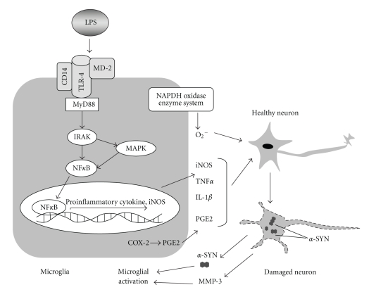
Parkinson's disease (PD) is a common neurodegenerative disorder characterized by the progressive loss of dopaminergic (DA) neurons in the substantia nigra. Although the exact cause of the dopaminergic neurodegeneration remains elusive, recent postmortem and experimental studies have revealed an essential role for neuroinflammation that is initiated and driven by activated microglial and infiltrated peripheral immune cells and their neurotoxic products (such as proinflammatory cytokines, reactive oxygen species, and nitric oxide) in the pathogenesis of PD. A bacterial endotoxin-based experimental model of PD has been established, representing a purely inflammation-driven animal model for the induction of nigrostriatal dopaminergic neurodegeneration. This model, by itself or together with genetic and toxin-based animal models, provides an important tool to delineate the precise mechanisms of neuroinflammation-mediated dopaminergic neuron loss. Here, we review the characteristics of this model and the contribution of neuroinflammatory processes, induced by the in vivo administration of bacterial endotoxin, to neurodegeneration. Furthermore, we summarize the recent experimental therapeutic strategies targeting endotoxin-induced neuroinflammation to elicit neuroprotection in the nigrostriatal dopaminergic system. The potential of the endotoxin-based PD model in the development of an early-stage specific diagnostic biomarker is also emphasized.
Figures
Similar articles
-
Protein kinase Cδ upregulation in microglia drives neuroinflammatory responses and dopaminergic neurodegeneration in experimental models of Parkinson's disease.Neurobiol Dis. 2016 Sep;93:96-114. doi: 10.1016/j.nbd.2016.04.008. Epub 2016 May 2. Neurobiol Dis. 2016. PMID: 27151770 Free PMC article.
-
Strategic selection of neuroinflammatory models in Parkinson's disease: evidence from experimental studies.CNS Neurol Disord Drug Targets. 2013 Aug;12(5):680-97. doi: 10.2174/18715273113129990059. CNS Neurol Disord Drug Targets. 2013. PMID: 23469840
-
Protective effects of endotoxin tolerance on peripheral lipopolysaccharide-induced neuroinflammation and dopaminergic neuronal injury.Immunopharmacol Immunotoxicol. 2022 Jun;44(3):326-337. doi: 10.1080/08923973.2022.2043900. Epub 2022 Mar 9. Immunopharmacol Immunotoxicol. 2022. PMID: 35260024
-
Environmental neurotoxicant-induced dopaminergic neurodegeneration: a potential link to impaired neuroinflammatory mechanisms.Pharmacol Ther. 2019 May;197:61-82. doi: 10.1016/j.pharmthera.2019.01.001. Epub 2019 Jan 22. Pharmacol Ther. 2019. PMID: 30677475 Free PMC article. Review.
-
Neuroinflammation in Parkinson's disease and its potential as therapeutic target.Transl Neurodegener. 2015 Oct 12;4:19. doi: 10.1186/s40035-015-0042-0. eCollection 2015. Transl Neurodegener. 2015. PMID: 26464797 Free PMC article. Review.
Cited by
-
Is Porphyromonas gingivalis involved in Parkinson's disease?Eur J Clin Microbiol Infect Dis. 2020 Nov;39(11):2013-2018. doi: 10.1007/s10096-020-03944-2. Epub 2020 Jun 21. Eur J Clin Microbiol Infect Dis. 2020. PMID: 32564247 Free PMC article. Review.
-
Elevated Percentage of CD3+ T-Cells and CD4+/CD8+ Ratios in Multiple System Atrophy Patients.Front Neurol. 2020 Jul 7;11:658. doi: 10.3389/fneur.2020.00658. eCollection 2020. Front Neurol. 2020. PMID: 32733370 Free PMC article.
-
The Tyrosine Phosphatase hPTPRβ Controls the Early Signals and Dopaminergic Cells Viability via the P2X7 Receptor.Int J Mol Sci. 2021 Nov 29;22(23):12936. doi: 10.3390/ijms222312936. Int J Mol Sci. 2021. PMID: 34884741 Free PMC article.
-
Protective effect of simvastatin on impaired intestine tight junction protein ZO-1 in a mouse model of Parkinson's disease.J Huazhong Univ Sci Technolog Med Sci. 2015 Dec;35(6):880-884. doi: 10.1007/s11596-015-1522-2. Epub 2015 Dec 16. J Huazhong Univ Sci Technolog Med Sci. 2015. PMID: 26670440
-
Lithium protects against paraquat neurotoxicity by NRF2 activation and miR-34a inhibition in SH-SY5Y cells.Front Cell Neurosci. 2015 May 28;9:209. doi: 10.3389/fncel.2015.00209. eCollection 2015. Front Cell Neurosci. 2015. PMID: 26074776 Free PMC article.
References
-
- Warner TT, Schapira AHV, Tatton , et al. Genetic and environmental factors in the cause of Parkinson’s disease. Annals of Neurology. 2003;53(3):S16–S25. - PubMed
-
- Lai BCL, Marion SA, Teschke K, Tsui JKC. Occupational and environmental risk factors for Parkinson’s disease. Parkinsonism and Related Disorders. 2002;8(5):297–309. - PubMed
-
- Lees AJ, Hardy J, Revesz T. Parkinson’s disease. The Lancet. 2009;373(9680):2055–2066. - PubMed
LinkOut - more resources
Full Text Sources
