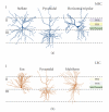
Selective vulnerability of neurons in layer II of the entorhinal cortex during aging and Alzheimer's disease
- PMID: 21331296
- PMCID: PMC3039218
- DOI: 10.1155/2010/108190
Selective vulnerability of neurons in layer II of the entorhinal cortex during aging and Alzheimer's disease
Abstract
All neurons are not created equal. Certain cell populations in specific brain regions are more susceptible to age-related changes that initiate regional and system-level dysfunction. In this respect, neurons in layer II of the entorhinal cortex are selectively vulnerable in aging and Alzheimer's disease (AD). This paper will cover several hypotheses that attempt to account for age-related alterations among this cell population. We consider whether specific developmental, anatomical, or biochemical features of neurons in layer II of the entorhinal cortex contribute to their particular sensitivity to aging and AD. The entorhinal cortex is a functionally heterogeneous environment, and we will also review data suggesting that, within the entorhinal cortex, there is subregional specificity for molecular alterations that may initiate cognitive decline. Taken together, the existing data point to a regional cascade in which entorhinal cortical alterations directly contribute to downstream changes in its primary afferent region, the hippocampus.
Figures


Similar articles
-
Activity disruption causes degeneration of entorhinal neurons in a mouse model of Alzheimer's circuit dysfunction.Elife. 2022 Dec 5;11:e83813. doi: 10.7554/eLife.83813. Elife. 2022. PMID: 36468693 Free PMC article.
-
Integrity of Neuronal Size in the Entorhinal Cortex Is a Biological Substrate of Exceptional Cognitive Aging.J Neurosci. 2022 Nov 9;42(45):8587-8594. doi: 10.1523/JNEUROSCI.0679-22.2022. Epub 2022 Sep 30. J Neurosci. 2022. PMID: 36180225 Free PMC article.
-
Microanatomical correlates of cognitive ability and decline: normal ageing, MCI, and Alzheimer's disease.Cereb Cortex. 2011 Aug;21(8):1870-8. doi: 10.1093/cercor/bhq264. Epub 2011 Jan 14. Cereb Cortex. 2011. PMID: 21239393
-
Molecular mechanisms of neurodegeneration in the entorhinal cortex that underlie its selective vulnerability during the pathogenesis of Alzheimer's disease.Biol Open. 2021 Jan 25;10(1):bio056796. doi: 10.1242/bio.056796. Biol Open. 2021. PMID: 33495355 Free PMC article. Review.
-
Should Alzheimer's disease be equated with human brain ageing? A maladaptive interaction between brain evolution and senescence.Ageing Res Rev. 2012 Jan;11(1):104-22. doi: 10.1016/j.arr.2011.06.004. Epub 2011 Jul 8. Ageing Res Rev. 2012. PMID: 21763787 Review.
Cited by
-
The Causal Role of Lipoxidative Damage in Mitochondrial Bioenergetic Dysfunction Linked to Alzheimer's Disease Pathology.Life (Basel). 2021 Apr 25;11(5):388. doi: 10.3390/life11050388. Life (Basel). 2021. PMID: 33923074 Free PMC article. Review.
-
A multi-hit hypothesis for an APOE4-dependent pathophysiological state.Eur J Neurosci. 2022 Nov;56(9):5476-5515. doi: 10.1111/ejn.15685. Epub 2022 Jun 6. Eur J Neurosci. 2022. PMID: 35510513 Free PMC article. Review.
-
Disrupted connectivity in the olfactory bulb-entorhinal cortex-dorsal hippocampus circuit is associated with recognition memory deficit in Alzheimer's disease model.Sci Rep. 2022 Mar 15;12(1):4394. doi: 10.1038/s41598-022-08528-y. Sci Rep. 2022. PMID: 35292712 Free PMC article.
-
Better stress coping associated with lower tau in amyloid-positive cognitively unimpaired older adults.Neurology. 2020 Apr 14;94(15):e1571-e1579. doi: 10.1212/WNL.0000000000008979. Epub 2020 Jan 21. Neurology. 2020. PMID: 31964689 Free PMC article.
-
Greater loss of object than spatial mnemonic discrimination in aged adults.Hippocampus. 2016 Apr;26(4):417-22. doi: 10.1002/hipo.22562. Epub 2016 Jan 18. Hippocampus. 2016. PMID: 26691235 Free PMC article.
References
Publication types
MeSH terms
Grants and funding
LinkOut - more resources
Full Text Sources
Medical

