

The Unfolded Protein Response, Degradation from Endoplasmic Reticulum and Cancer
- PMID: 21331300
- PMCID: PMC3039444
- DOI: 10.1177/1947601910383011
The Unfolded Protein Response, Degradation from Endoplasmic Reticulum and Cancer
Abstract
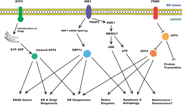
The endoplasmic reticulum (ER) is an essential organelle involved in many cellular functions including protein folding and secretion, lipid biosynthesis and calcium homeostasis. Proteins destined for the cell surface or for secretion are made in the ER, where they are folded and assembled into multi-subunit complexes. The ER plays a vital role in cellular protein quality control by extracting and degrading proteins that are not correctly folded or assembled into native complexes. This process, known as ER-associated degradation (ERAD), ensures that only properly folded and assembled proteins are transported to their final destinations. Besides its role in protein folding and transport in the secretory pathway, the ER regulates the biosynthesis of cholesterol and other membrane lipids. ERAD is an important means to ensure that levels of the responsible enzymes are appropriately maintained. The ER is also a major organelle for oxygen and nutrient sensing as cells adapt to their microenvironment. Stresses that disrupt ER function leads to accumulation of unfolded proteins in the ER, a condition known as ER stress. Cells adapt to ER stress by activating an integrated signal transduction pathway called the unfolded protein response (UPR) (1). The UPR represents a survival response by the cells to restore ER homeostasis. If ER stress persists, cells activate mechanisms that result in cell death. Chronic ER stress is increasingly being recognized as a factor in many human diseases such as diabetes, neurodegenerative disorders and cancer. In this review we discuss the roles of the UPR and ERAD in cancer and suggest directions for future research.
Conflict of interest statement
The author(s) declared no potential conflicts of interest with respect to the authorship and/or publication of this article.
Figures
References
-
- Ron D, Walter P. Signal integration in the endoplasmic reticulum unfolded protein response. Nat Rev Mol Cell Biol. 2007;8:519-29 - PubMed
-
- Bertolotti A, Zhang Y, Hendershot LM, Harding HP, Ron D. Dynamic interaction of BiP and ER stress transducers in the unfolded-protein response. Nat Cell Biol. 2000;2:326-32 - PubMed
-
- Shen J, Chen X, Hendershot L, Prywes R. ER stress regulation of ATF6 localization by dissociation of BiP/GRP78 binding and unmasking of Golgi localization signals. Dev Cell. 2002;3:99-111 - PubMed
-
- Shen J, Prywes R. Dependence of site-2 protease cleavage of ATF6 on prior site-1 protease digestion is determined by the size of the luminal domain of ATF6. J Biol Chem. 2004;279:43046-51 - PubMed
-
- Wang Y, Shen J, Arenzana N, Tirasophon W, Kaufman RJ, Prywes R. Activation of ATF6 and an ATF6 DNA binding site by the endoplasmic reticulum stress response. J Biol Chem. 2000;275:27013-20 - PubMed
Grants and funding
LinkOut - more resources
Full Text Sources
Other Literature Sources