

doi: 10.1155/2010/491928.
Epub 2011 Feb 9.
Neural pathways in allergic inflammation
Affiliations
- PMID: 21331366
- PMCID: PMC3038426
- DOI: 10.1155/2010/491928
Item in Clipboard
Neural pathways in allergic inflammation
J Allergy (Cairo).
2010.
Abstract
Allergy is on the rise worldwide. Asthma, food allergy, dermatitis, and systemic anaphylaxis are amongst the most common allergic diseases. The association between allergy and altered behavior patterns has long been recognized. The molecular and cellular pathways in the bidirectional interactions of nervous and immune systems are now starting to be elucidated. In this paper, we outline the consequences of allergic diseases, especially food allergy and asthma, on behavior and neural activity and on the neural modulation of allergic responses.
Figures
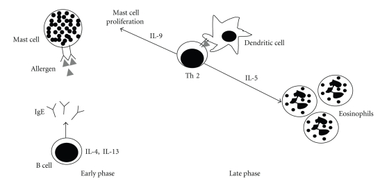
Early phase and late phase of allergic hypersensitivity. Upon allergen challenge, sensitized individuals can present two distinct phases: the early phase, which is characterized by mast cell degranulation and release of inflammatory mediators triggered by cross-linking IgE antibodies present on mast cells membranes, and the late phase, that is characterized by the infiltration of Th2 cells that interact with dendritic cells releasing type 2 cytokines responsible for tissue mast cell proliferation and eosinophil recruitment.
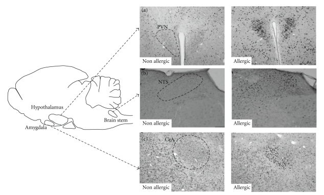
Activation of specific brain areas by c-fos expression. Representative brain coronal sections of nonallergic (nonsensitized) and allergic (OVA-sensitized) mice after (oral or nasal) challenge with OVA. Fos staining in neurons of (a) the paraventricular nucleus of the hypothalamus (PVN), (b) nucleus of the tract solitary (NTS), and (c) central nucleus of the amygdala (CeA). Adapted from Basso et al. 2004 [36] and Costa-Pinto et al. [47].
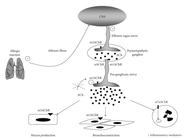
The cholinergic pathways in allergic lung. During allergic reactions, the inflammatory mediators released in the tissue activate the sensory afferent fibers, which convey information to the CNS. The CNS sends information back to the inflammatory site by increasing ACh release from efferent vagus nerve. The neurotransmission in the parasympathetic ganglia is mediated by acetylcholine (ACh) via nicotinic (nAChR) or type 1 muscarinic (m1AChR) receptors. The stimulus generated induces ACh release in the pos ganglionic nerve fiber endings. Type 2 muscarinic receptors (m2AChRs) are autoinhibitory, and the dysfunction of this receptor, observed in allergic asthma, induces increased release of ACh. Increased ACh results in augmented mucus secretion via m3AchR expressed in the glandular epithelium, increased airway smooth muscle contraction (bronchoconstriction) via m3AchR expressed in muscle cells, and decreased inflammatory mediators production via a7nAChR receptor expressed on immune cells.
Similar articles
-
Advances and novel developments in mechanisms of allergic inflammation.Allergy. 2020 Dec;75(12):3100-3111. doi: 10.1111/all.14632. Epub 2020 Nov 4. Allergy. 2020. PMID: 33068299 Review.
-
Good allergy practice--standards of care for providers and purchasers of allergy services within the National Health Service. Royal College of Physicians and Royal College of Pathologists.Clin Exp Allergy. 1995 Jul;25(7):586-95. Clin Exp Allergy. 1995. PMID: 8521176
-
microRNA and Allergy.Adv Exp Med Biol. 2015;888:331-52. doi: 10.1007/978-3-319-22671-2_17. Adv Exp Med Biol. 2015. PMID: 26663191
-
Cholinergic Modulation of Type 2 Immune Responses.Front Immunol. 2017 Dec 19;8:1873. doi: 10.3389/fimmu.2017.01873. eCollection 2017. Front Immunol. 2017. PMID: 29312347 Free PMC article. Review.
-
What Can Parasites Tell Us About the Pathogenesis and Treatment of Asthma and Allergic Diseases.Front Immunol. 2020 Sep 11;11:2106. doi: 10.3389/fimmu.2020.02106. eCollection 2020. Front Immunol. 2020. PMID: 33013887 Free PMC article. Review.
Cited by
-
Allergic host defences.Nature. 2012 Apr 25;484(7395):465-72. doi: 10.1038/nature11047. Nature. 2012. PMID: 22538607 Free PMC article.
-
Acute oral ethanol exposure triggers asthma in cockroach allergen-sensitized mice.Am J Pathol. 2012 Sep;181(3):845-57. doi: 10.1016/j.ajpath.2012.05.020. Epub 2012 Jul 13. Am J Pathol. 2012. PMID: 22796441 Free PMC article.
-
Heart Rate Variability in Subjects with Severe Allergic Background Undergoing COVID-19 Vaccination.Vaccines (Basel). 2023 Mar 1;11(3):567. doi: 10.3390/vaccines11030567. Vaccines (Basel). 2023. PMID: 36992151 Free PMC article.
-
Food components and the immune system: from tonic agents to allergens.Front Immunol. 2013 May 17;4:102. doi: 10.3389/fimmu.2013.00102. eCollection 2013. Front Immunol. 2013. PMID: 23730302 Free PMC article.
-
Dissociation between sickness behavior and emotionality during lipopolysaccharide challenge in lymphocyte deficient Rag2(-/-) mice.Behav Brain Res. 2015 Feb 1;278:74-82. doi: 10.1016/j.bbr.2014.09.030. Epub 2014 Sep 22. Behav Brain Res. 2015. PMID: 25257108 Free PMC article.
References
-
- Ono SJ. Molecular genetics of allergic diseases. Annual Review of Immunology. 2000;18:347–366. - PubMed
-
- Gould HJ, Sutton BJ. IgE in allergy and asthma today. Nature Reviews Immunology. 2008;8(3):205–217. - PubMed
-
- de Sousa Mucida D, de Castro Keller A, Fernvik EC, Russo M. Unconventional strategies for the suppression of allergic asthma. Current Drug Targets. Inflammation and Allergy. 2003;2(2):187–195. - PubMed
-
- Maddox L, Schwartz DA. The pathophysiology of asthma. Annual Review of Medicine. 2002;53:477–498. - PubMed
LinkOut - more resources
Full Text Sources
